




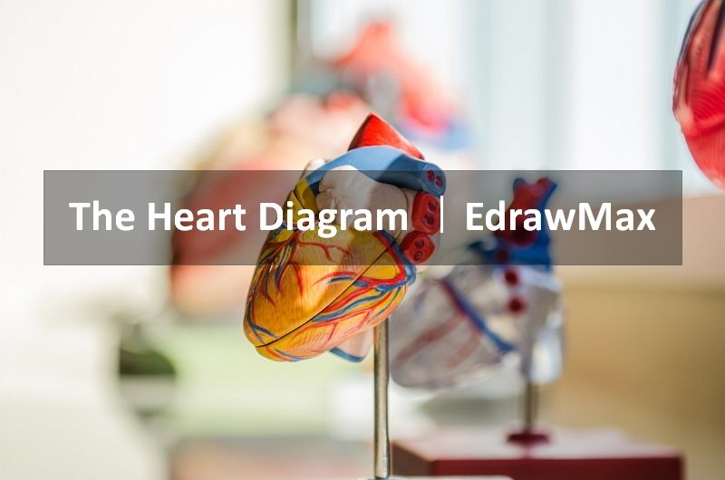
The human heart is a muscular pump with four chambers: the right atrium, right ventricle, left atrium, and left ventricle. It sits between the lungs, behind the sternum, and slightly left of the body's midline. A protective sac called the pericardium surrounds it. For a clinical overview of the same structures, see Cleveland Clinic's heart anatomy guide.
A clear heart diagram should show the four chambers, four valves, major blood vessels, and the direction of blood flow. The right side sends oxygen-poor blood to the lungs; the left side receives oxygen-rich blood from the lungs and pumps it through the aorta to the body.
Heart blood-flow path at a glance: body → venae cavae → right atrium → tricuspid valve → right ventricle → pulmonary valve → pulmonary arteries → lungs → pulmonary veins → left atrium → mitral valve → left ventricle → aortic valve → aorta → body. This sequence is the fastest way to read a labeled heart diagram without treating each label as an isolated part.
In this article
For a labeled heart diagram, focus on three groups: the four chambers, the four valves, and the major vessels that carry blood into and out of the heart. Reading those groups together makes the direction of blood flow much easier to follow.
1. What Does the Human Heart Look Like?
The heart is about the size of a person's fist and is made primarily of cardiac muscle. A wall called the septum separates the right and left sides. The upper chambers are the atria, which receive blood, and the lower chambers are the ventricles, which pump blood out of the heart.
At a glance: The human heart has four chambers and four main valves. Blood returns from the body to the right side of the heart, goes to the lungs for oxygen, returns to the left side, and is pumped through the aorta to the body; the valves keep this flow moving in one direction.
| Structure | Main function |
|---|---|
| Right atrium | Receives oxygen-poor blood returning from the body |
| Right ventricle | Pumps blood to the lungs through the pulmonary artery |
| Left atrium | Receives oxygen-rich blood from the lungs through the pulmonary veins |
| Left ventricle | Pumps oxygen-rich blood into the aorta and systemic circulation |
| Septum | Separates the right and left sides of the heart |
| Pericardium | Protective sac surrounding the heart |
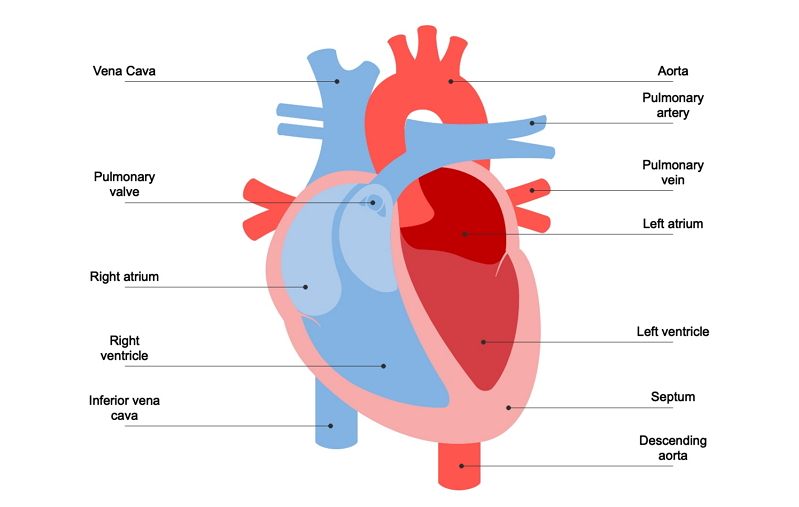
2. How Does Blood Flow Through the Heart?
Blood follows a repeating one-way route through the chambers, valves, lungs, and body. The heart valves open to let blood move forward and close to reduce backward flow.
- Oxygen-poor blood from the body returns through the superior and inferior venae cavae to the right atrium.
- It passes through the tricuspid valve into the right ventricle.
- The right ventricle pumps it through the pulmonary valve into the pulmonary arteries.
- The pulmonary arteries carry the blood to the lungs, where gas exchange adds oxygen and removes carbon dioxide.
- Oxygen-rich blood returns through the pulmonary veins to the left atrium.
- It passes through the mitral valve into the left ventricle.
- The left ventricle pumps blood through the aortic valve into the aorta.
- The aorta distributes oxygen-rich blood to the body before it returns to the right side of the heart.
Key distinction: the pulmonary arteries carry oxygen-poor blood from the right ventricle to the lungs, while the aorta carries oxygen-rich blood from the left ventricle to the body. Keeping these two routes separate makes labeled heart diagrams easier to read correctly.
3. What Do the Heart Valves and Major Blood Vessels Do?
| Part | Direction / role |
|---|---|
| Tricuspid valve | Right atrium → right ventricle |
| Pulmonary valve | Right ventricle → pulmonary artery |
| Mitral valve | Left atrium → left ventricle |
| Aortic valve | Left ventricle → aorta |
| Venae cavae | Return oxygen-poor blood from the body to the right atrium |
| Pulmonary arteries | Carry oxygen-poor blood from the right ventricle to the lungs |
| Pulmonary veins | Carry oxygen-rich blood from the lungs to the left atrium |
| Aorta | Carries oxygen-rich blood from the left ventricle to the body |
| Coronary arteries | Supply oxygen-rich blood to the heart muscle itself |
The right and left coronary arteries arise from the aorta and supply the myocardium. It is incorrect to say that the left coronary artery carries blood to the right atrium and right ventricle as its primary function.
4. How Do You Draw and Label a Heart Diagram?
For an educational heart diagram, focus on correct relationships and flow direction before adding decorative detail.
- Draw the four chambers. Place the atria above the ventricles and separate the right and left sides with the septum.
- Add the four valves. Position each valve between its chamber pair or outflow vessel.
- Add the major vessels. Include the venae cavae, pulmonary arteries, pulmonary veins, and aorta.
- Add arrows for blood flow. Use a continuous route from body → right heart → lungs → left heart → body.
- Label oxygen-poor and oxygen-rich blood carefully. Remember that pulmonary arteries carry oxygen-poor blood and pulmonary veins carry oxygen-rich blood.

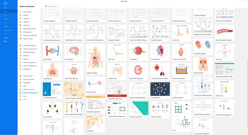
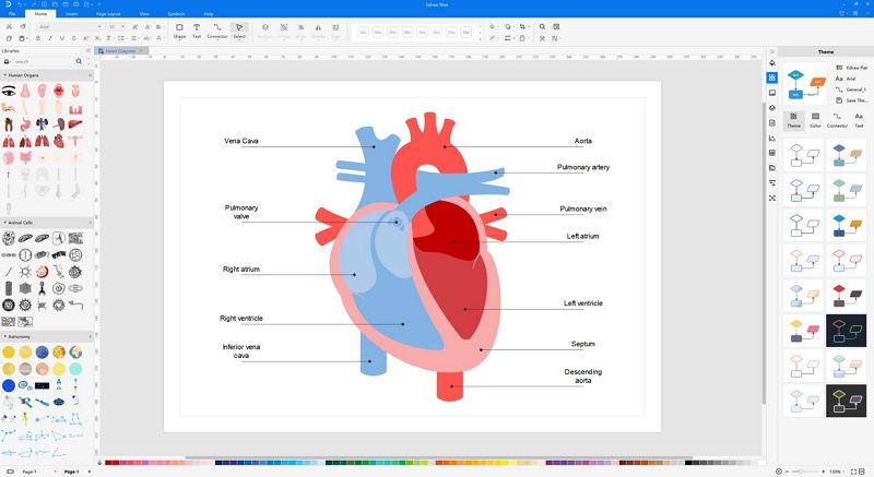
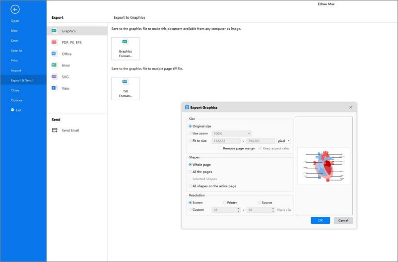
How to Create a Heart Diagram in EdrawMax
Use a science or anatomy template as a starting point, then label the chambers, valves, and vessels and add directional arrows. The main quality check is anatomical consistency: every label and arrow should match the actual direction of blood flow.
 Get 500 free AI tokens
5. FAQs About the Human Heart Diagram.
What should a labeled heart diagram include?
At minimum, a study diagram should label the four chambers, the tricuspid, pulmonary, mitral, and aortic valves, the venae cavae, pulmonary arteries, pulmonary veins, and aorta. Arrows showing blood-flow direction make the relationship between these structures much easier to understand.
How many chambers does the human heart have?
Four: the right atrium, right ventricle, left atrium, and left ventricle.
Which side of the heart pumps blood to the lungs?
The right ventricle pumps oxygen-poor blood through the pulmonary valve and pulmonary arteries to the lungs.
Which chamber pumps blood to the body?
The left ventricle pumps oxygen-rich blood through the aortic valve into the aorta and systemic circulation.
How many valves are in the heart?
A normal heart has four main valves: tricuspid, pulmonary, mitral, and aortic.
What is the difference between pulmonary arteries and pulmonary veins?
Pulmonary arteries carry oxygen-poor blood from the heart to the lungs. Pulmonary veins carry oxygen-rich blood from the lungs back to the heart.
References: American Heart Association: Heart Valves and Circulation and Cleveland Clinic: Heart Chambers.
AI Diagram Generator
Enter your prompt. Upload files if needed. Generate diagrams, charts, or slides instantly.





 (AI & Web)