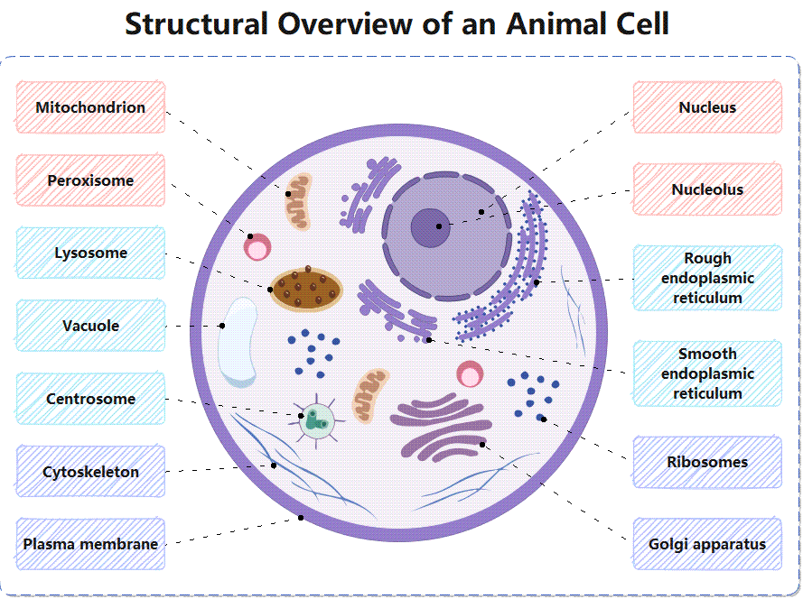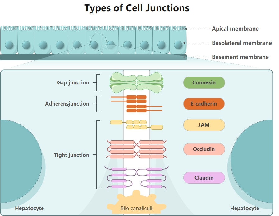
About this Cell Junction Diagram template
This cell junction diagram template provides a clear visual of how cells connect and communicate. It highlights the main types of junctions found in biological tissues. You can use this to study or teach complex cellular structures easily.
Tight Junctions
Tight junctions act as a seal to prevent fluids from leaking between cells. They are crucial for maintaining barriers in organs like the gut or liver. These junctions ensure that molecules move through controlled paths.
- JAM (Junctional Adhesion Molecules)
- Occludin
- Claudin
Adherens Junctions
Adherens junctions provide strong mechanical attachments between adjacent cells. They help tissues resist stretching and remain stable under physical stress. These connections link the internal skeletons of cells together to form a very sturdy unit.
- E-cadherin
- Actin filaments
Gap Junctions
Gap junctions serve as communication channels between cells. They allow small molecules and ions to pass directly from one cell to another. This fast sharing is vital for coordinating activities like heart muscle contractions.
- Connexin proteins
- Intercellular channels
FAQs about this Template
-
What is the primary function of tight junctions in the body?
Tight junctions serve as the body's primary seal to prevent leakage between cells. They create a waterproof barrier that forces nutrients and fluids to pass through cells instead of around them. This is essential in organs like the bladder and stomach. By holding cells closely together, they maintain the distinct internal environments needed for specialized organ functions to work correctly.
-
How do gap junctions facilitate cell-to-cell communication?
Gap junctions act like tiny tunnels or bridges connecting the cytoplasm of neighboring cells. They are made of proteins called connexins that form a passage. This allows ions and small molecules to flow freely between neighbors. Such direct contact is crucial for synchronized movements, like the rhythmic beating of the heart or the rapid spread of electrical signals in the nervous system.
-
Why are adherens junctions important for tissue stability?
Adherens junctions are like biological velcro that keeps cells stuck together during physical movement. They connect to the cell's cytoskeleton, providing deep structural support. This prevents tissues from tearing apart when they are pulled or stretched. You will find them heavily used in skin and muscle tissues, where maintaining a strong physical bond between many individual cells is absolutely necessary.