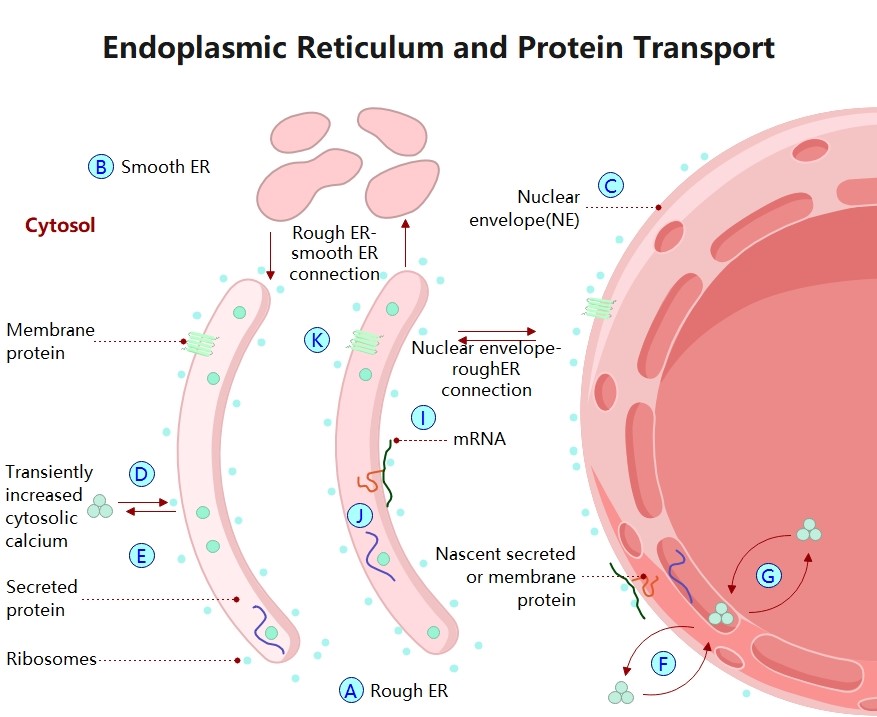
About this Endoplasmic Reticulum Structure
This biological diagram illustrates the complex membrane system of the endoplasmic reticulum. It helps students visualize the physical differences between rough and smooth regions, highlighting how their unique shapes and surface features facilitate distinct cellular tasks like protein folding and detoxification.
Rough Endoplasmic Reticulum (RER)
The rough ER is identifiable by the ribosomes attached to its surface, giving it a studded appearance. It focuses on the synthesis and folding of proteins that are destined for secretion or membrane integration.
- Surface-bound ribosomes
- Flattened sacs called cisternae
- Primary site of protein synthesis
- Directly connected to nuclear envelope
Smooth Endoplasmic Reticulum (SER)
Smooth ER lacks ribosomes and primarily consists of a branching network of tubules. It is responsible for creating lipids and steroid hormones, while also assisting in the detoxification of various harmful chemical substances.
- Tubular membrane network
- Lipid and steroid production
- Drug and toxin metabolism
- Calcium ion storage site
Cisternae and Lumen
The ER consists of membrane-enclosed sacs called cisternae, which surround an internal space known as the lumen. This enclosed environment allows for specific enzymatic reactions and provides the space needed for proper protein folding.
- Internal ER lumen space
- Folded cisternal membranes
- High surface area for reactions
- Transport vesicle formation site
FAQs about this Template
-
Why is it called "rough" and "smooth" endoplasmic reticulum?
The names describe the appearance of these organelles under a microscope. The rough ER is covered with tiny ribosomes, which look like bumps or grains on its surface. These ribosomes are essential for building proteins. In contrast, the smooth ER lacks these ribosomes, resulting in a sleek, tubular appearance that is primarily used for producing fats and processing metabolic waste products.
-
How does the ER communicate with the Golgi apparatus?
The endoplasmic reticulum and Golgi apparatus work together through a system of transport vesicles. Once the ER synthesizes proteins or lipids, it packages them into small, membrane-bound sacs. These vesicles then bud off from the ER and travel to the Golgi body. There, the molecules are further refined, tagged with chemical signals, and finally shipped to their specific cellular or extracellular destinations.
-
What is the significance of the ER's connection to the nucleus?
The ER membrane is physically continuous with the outer layer of the nuclear envelope. This connection is vital because it allows messenger RNA to move directly from the nucleus to the rough ER's ribosomes. This immediate proximity ensures that the genetic code can be translated into proteins quickly and efficiently, minimizing the distance molecules must travel within the cell's crowded interior.