About this viral infection example
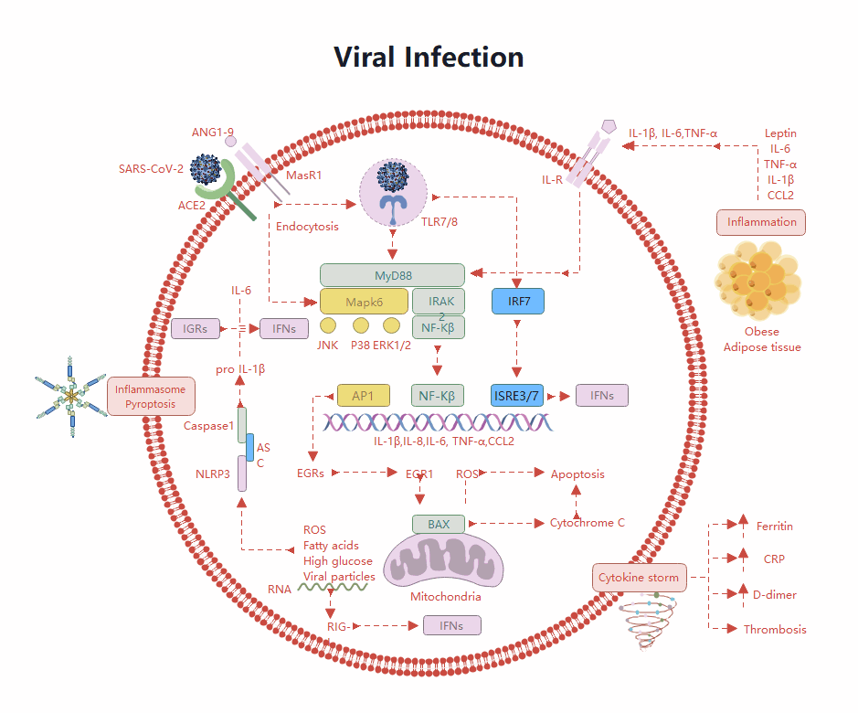
This template illustrates the complex biological process of a viral infection at the cellular level. It specifically maps out how a virus enters a host cell and triggers various immune system responses. Use this diagram to study molecular pathways and inflammatory reactions clearly.
Viral Entry and Endocytosis
The virus begins its attack by attaching to specific receptors on the cell surface. This process allows the viral particles to enter the cell through endocytosis. Once inside, the virus releases its genetic material to start the infection.
- SARS-CoV-2 binding to ACE2
- ACE2 and MasR1 receptors
- Endocytosis mechanism
- TLR7/8 sensing
Cellular Immune Signaling
After the virus enters, the cell activates several signaling pathways to fight the invader. These pathways involve proteins like MyD88 and NF-kappaB, which travel to the nucleus. They trigger the production of defense molecules and inflammatory markers.
- MyD88 and MAPK signaling
- NF-kappaB and IRF7 activation
- Interferon (IFNs) production
- Transcription factors AP1 and ISRE3/7
Cell Death and Cytokine Storm
Severe infections can lead to uncontrolled immune responses known as a cytokine storm. This stage involves programmed cell death and significant tissue damage. The body produces excess proteins that can cause dangerous blood clots or systemic inflammation.
- Inflammasome and Pyroptosis
- Mitochondrial BAX and Cytochrome C
- Cytokine storm and Thrombosis
- Ferritin and CRP markers
FAQs about this Template
-
How does the host cell recognize a viral infection?
Host cells use specialized receptors called Pattern Recognition Receptors, such as TLR7 and TLR8, to detect viral components like RNA. Once these sensors identify the virus, they trigger signaling molecules like MyD88. This process initiates a cascade that activates transcription factors, leading to the production of interferons and cytokines to stop the virus from spreading further.
-
What role do mitochondria play in the immune response?
Mitochondria act as vital hubs for immune signaling and cellular stress detection during an infection. They can release reactive oxygen species and proteins like Cytochrome C to trigger apoptosis, or programmed cell death. This prevents the virus from hijacking the cell's machinery for replication, effectively sacrificing the infected cell to protect the rest of the host organism.
-
What is a cytokine storm and why is it dangerous?
A cytokine storm is an overactive immune response where the body releases too many inflammatory proteins, such as IL-6 and TNF-alpha, into the blood too quickly. This can lead to widespread tissue damage, organ failure, and blood clotting issues like thrombosis. It represents a breakdown in immune regulation where the body's defense system begins harming itself.