


Leaves are one of the most important organs of a plant, acting as natural food-making factories. Using sunlight, air, and water, a leaf produces food through the process of photosynthesis, supporting not only itself but the entire plant. To truly understand how this process works, it is essential to study the internal and external structure of a leaf.
In this complete anatomy guide, we will explore the plant leaf structure in a simple and well-organized way using labelled diagrams. You will learn about the main components of a leaf, including the epidermis, mesophyll tissues, vascular bundles, and stomata, along with their specific functions.
This guide also explains how stomata open and close, the differences between simple and compound leaves, and a detailed monocot vs dicot leaf anatomy comparison. In addition, you will find an easy leaf cross-section drawing tutorial and free diagram templates to help students and teachers visualize leaf anatomy more effectively.
Whether you are a biology student, teacher, or beginner, this guide will help you clearly understand the structure and function of plant leaves.
In this article
What Are the Main Parts of a Leaf?
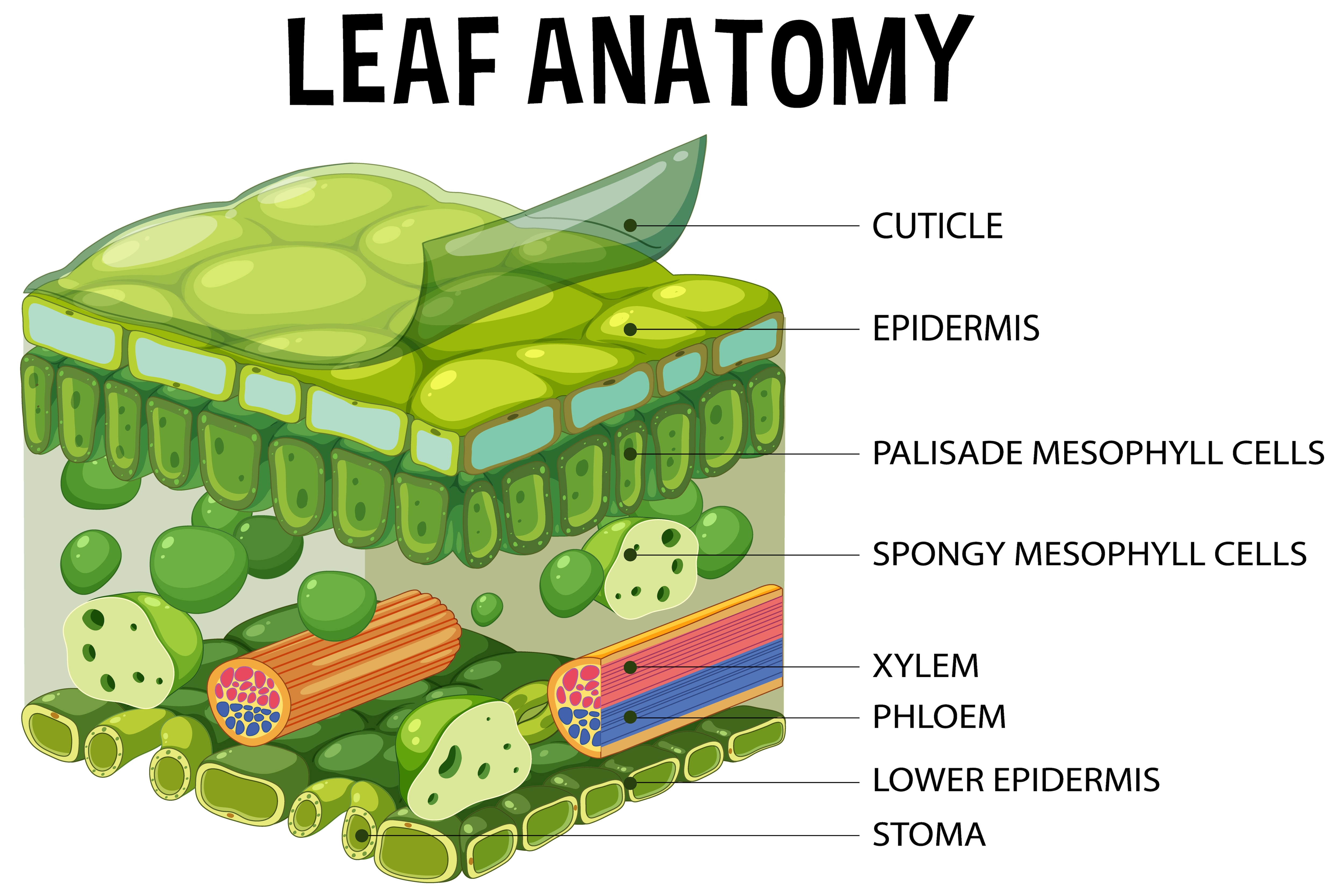
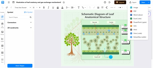
The main components of a leaf structure are the mesophyll, vascular bundles, and epidermis.
Epidermis: The Protective Outer Layer
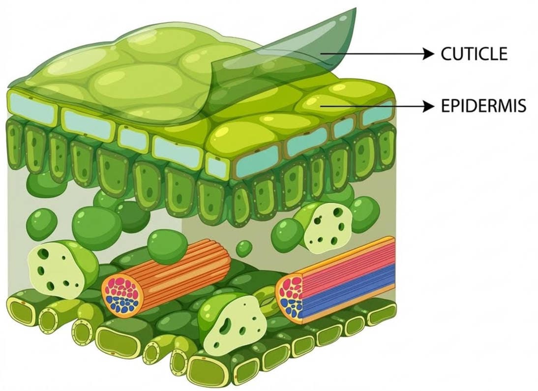
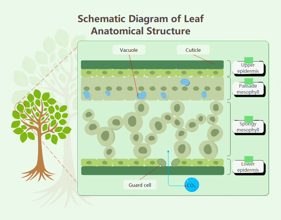
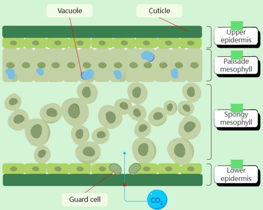
The epidermis is the first layer on the outer side of the leaf. It protects the plant from dryness and damage from external surroundings. It consists of two layers: the upper epidermis and the lower epidermis.
In most plants, stomata are present in the lower epidermis and perform the function of gas exchange. The epidermis also functions as a semipermeable membrane. It only allows necessary materials to pass through the leaf. Another waxy layer called the cuticle covers it, preventing water loss.
Mesophyll: The Photosynthesis Powerhouse
The mesophyll is present inside the upper and lower epidermis. It is the main part of a leaf where photosynthesis occurs. The mesophyll has two components: the palisade mesophyll and the spongy mesophyll.
The palisade mesophyll contains chloroplasts that absorb sunlight. The spongy mesophyll contains air spaces that help in gas exchange. Xylem transports water and minerals from the roots to the mesophyll. Then the process of photosynthesis makes food.
Vascular Bundles: Xylem and Phloem Transport
Vascular bundles work as the transport system for the plants. They are present in the veins of a leaf near the mesophyll and transport food, minerals, and water to all parts of a plant. There are two types of vascular bundles:
- Xylem: conducts water and minerals from the soil to the leaves
- Phloem: transports the food from the leaves to other parts of a plant
How Do Stomata Work?
The stomata are the tiny pores on the surface of a leaf. They exchange carbon dioxide, oxygen, and water vapor.
Guard Cells and Gas Exchange Mechanism
Two guard cells surround each stomata and help to exchange carbon dioxide, oxygen, and water. In sunlight, potassium ions accumulate in large amounts in guard cells, causing them to become rigid.
The stomatal pore opens due to this pressure. At night, guard cells lose potassium ions because sunlight is absent. This loss of potassium ions makes guard cells flaccid. In this way, stomata are closed.
Stomatal Opening and Closing: Environmental Control
The opening and closing of stomata happen in response to environmental conditions. In the daytime, stomata open and absorb carbon dioxide from the air for photosynthesis. Therefore, leaves prepare food in the sunlight.
At night, stomata close because there is no sunlight to prepare food. Similarly, stomata close in hot, dry conditions to conserve water. This process of stomatal opening and closing is crucial for respiration and preventing water loss.
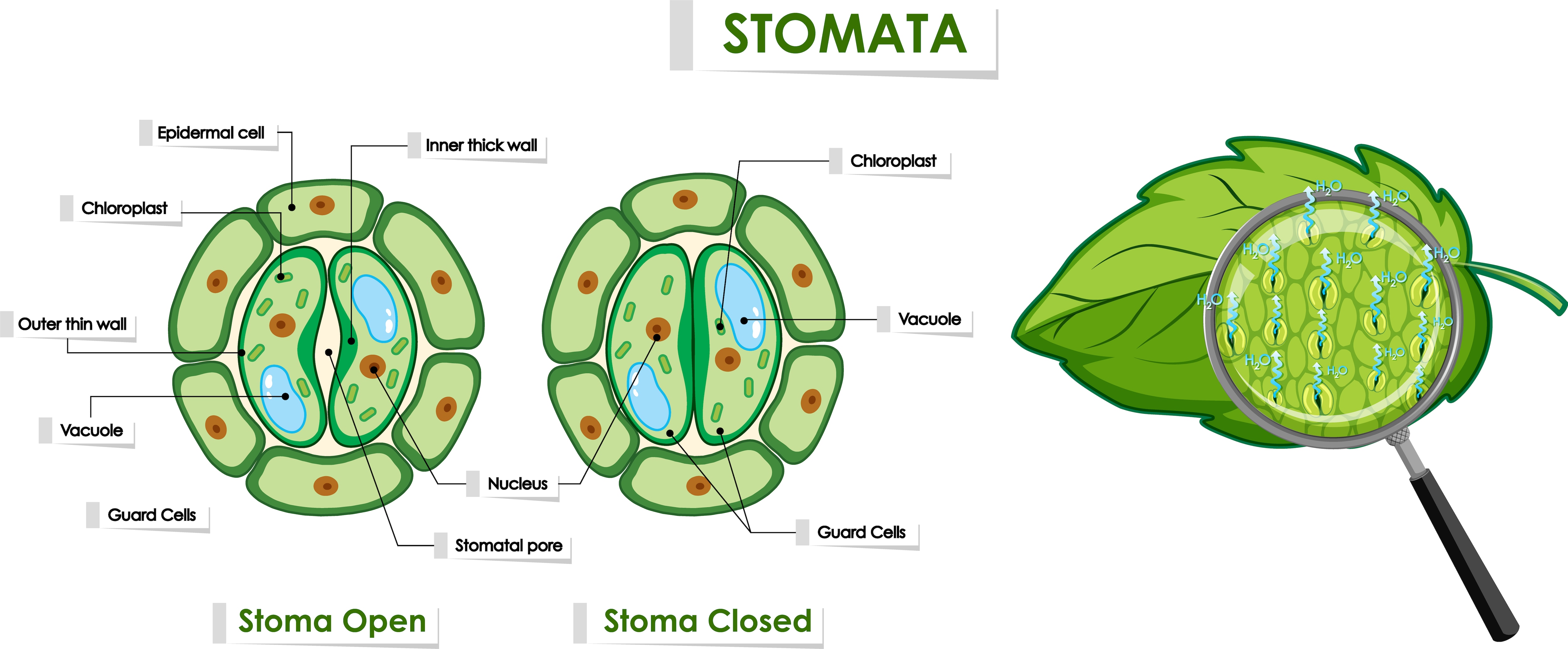
Visualizing Stomatal Function Through Diagrams
Because stomatal movement involves changes in cell shape and internal pressure, diagrams are especially helpful for understanding this process.
A clear, labeled diagram can show the position of guard cells, the stomatal pore, and the differences between open and closed states. Drawing or studying such diagrams helps students visualize these microscopic structures more effectively.
To better understand leaf anatomy, students often create their own diagrams. Digital diagramming tools can simplify this process by providing basic shapes and biology-related symbols. These tools allow learners to focus on structure and labeling rather than drawing accuracy.
EdrawMax is an advanced diagramming tool used for biological diagrams such as leaf anatomy and stomatal structure. It offers a wide range of ready-made templates, symbols, and shapes that make drawing scientific diagrams simple and accurate. Its user-friendly features make it especially useful for students and teachers.
How to Make a Leaf Structure Diagram with EdrawMax
Here is a step-by-step tutorial to draw a leaf structure diagram using EdrawMax:
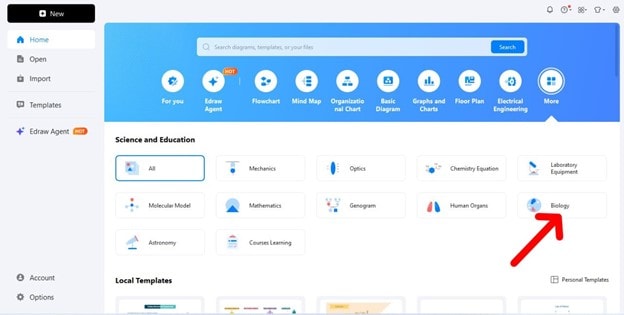
Step1 Download EdrawMax
Download EdrawMax or try it online. Navigate to Biology from the Science and Education section
Step2 Search Leaf Structure Templates
Search "leaf structure diagram templates" to find a template and open it for editing.
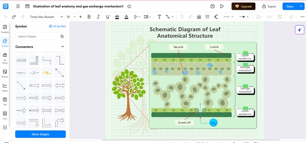
Step3 Edit the Template
You can now see how to edit each part by clicking. The colors and size of components can be changed. You can also label different parts, such as the epidermis and palisade mesophyll, and spongy mesophyll.
Moreover, you can choose other leaf structure templates, such as the one shown below.
Step4 Export and Share Diagram
After finishing the editing, you can export the diagram as a PDF, SVG, MS Office File, or JPG. Now you can share the diagram with your friends, students, or teachers to clearly understand the leaf structure.
In fact, you can export the final result as a visually appealing GIF image as shown below:
Different Types of Leaf Structures
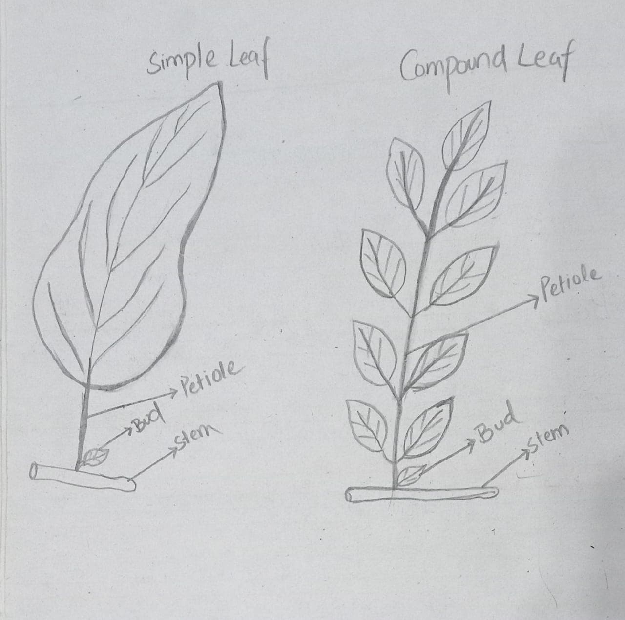
Simple vs Compound Leaves
Leaves are divided into two types based on the structure of their leaf blade. Simple leaves have a single, undivided blade attached to the stem by a petiole. For example, mango, pear, and maple have simple leaves.
Compound leaves have a blade that divides into multiple units, called leaflets. For example, rose, neem, and pea have compound leaves. The differences between simple and compound leaves are presented in the table below:
| Sr. No. | Simple Leaves | Compound Leaves |
| Blade division | There is a single, undivided blade | The blade is divided into smaller units called leaflets. |
| Leaf arrangement | Follows acropetal succession, where older leaves are at the bottom, and new leaves grow upwards | The leaflets do not follow acropetal succession |
| Attachment to stem | A single petiole attaches a simple leaf to a stem. | A petiole attaches all the leaflets of a compound leaf. |
Monocot vs Dicot Leaf Anatomy Comparison
There are two groups of plants based on the number of cotyledons in their seed: monocots and dicots. Both have different leaf structures. The table explains the key differences in the anatomy of monocots and dicots:
| Sr. No. | Monocots | Dicots |
| Leaf shape and venation | Long and narrow leaves with parallel venation | Broader leaves with reticulate venation |
| Structure of the upper and lower epidermis | Not much difference; both are the same. | Clear difference |
| Mesophyll Differentiation | Not clearly differentiated | Differentiated into palisade and spongy mesophyll tissues |
| Distribution of stomata | Present on both the upper and lower epidermis | Mostly present on the lower epidermis |
| Number and position of vascular bundles | More vascular bundles are scattered | Fewer numbers of vascular bundles, and they are arranged in the proper position |
| Examples | Wheat, maize, rice | Sunflower, pea, cotton |
Bonus Part: How to Draw a Leaf Cross-Section?
The cross-section of a leaf shows its anatomy consisting of upper and lower epidermis, mesophyll tissues, vascular bundles, and stomata. Students may feel that drawing a cross-section diagram of a leaf is hard, but it is not. You can draw and label this diagram just by following this tutorial.
Drawing Upper and Lower Epidermis Layers
- Start the diagram by drawing two horizontal thin lines to show the upper and lower epidermis.
- Now, draw a wavy pattern on both lines and make layers of rectangular epidermal cells. These rectangular cells must be closely packed.
- Make sure to leave some spaces on the lower epidermis to show stomata.
- Draw an extra layer on the upper epidermal layer to show the cuticle protection.
- Color the epidermal layer and cuticle layer to enhance visibility and clarity.
Illustrating Palisade and Spongy Mesophyll
- The mesophyll tissue lies between the two epidermal layers.
- To illustrate the palisade mesophyll, Draw elongated rod-shaped structures below the upper epidermis. Make thick boundaries around these structures.
- Chloroplasts are present in these cells for photosynthesis. So, add some dark green dots to show chloroplasts.
- Next, draw some irregular spongy-shaped structures between the palisade mesophyll and lower epidermis. Draw them with large gaps to show spongy mesophyll tissues.
- Also, draw oval or bean-shaped structures for vascular bundles near the spongy mesophyll.
Showing Stomata Structure in Plant Anatomy
- Stomata are opening pores for respiration and gas exchange. Guard cells control their opening and closing. To understand stomata anatomy, you need to know the structure of guard cells and stomata.
- Draw two kidney-shaped structures facing each other to show guard cells
- To show open stomata, leave some space between them. In this situation, guard cells are filled with water and potassium ions.
- To show closed stomata, there will be no space between the guard cells, and the guard cells will be flaccid.
AI Diagram Generator
Enter your prompt. Upload files if needed. Generate diagrams, charts, or slides instantly.