


Biology is incomplete without diagrams. These diagrams help us understand the complex processes that occur in our cells. The endomembrane system is one of the complex systems happening in our cells.
The purpose of this guide is to learn how the endomembrane system diagram can be designed most easily. So without further ado, let’s get started.
In this article
What Is the Endomembrane System?
The endomembrane system is one of the most intricate and vital organizational systems in a eukaryotic cell. It represents a complex network of internal membranes that work in coordination to modify, package, and transport proteins and lipids throughout the cell.
Components and Functions
The endomembrane system consists of several organelles that are membrane-bound, especially those whose membranes are directly connected or exchange materials. Here are components of the endomembrane system and their functions:
- The nuclear envelope: it surrounds the nucleus and serves as the starting point of the whole system. It is connected with the ER and allows mRNA to exit the nucleus.
- Endoplasmic Reticulum: It is a vast network of tubules and flattened sacs. The rough ER is responsible for protein synthesis and modification, while the smooth ER deals with lipid synthesis.
- The Golgi apparatus: it is usually called the post office of the cell. It receives materials from the ER, modifies, sorts, packages, and tags them for delivery within the cell or outside the cell.
- Plasma membrane: lipids and proteins are part of the plasma membrane, which are synthesized and processed through the ER and Golgi and transferred via vesicles to be integrated.
- Vesicles and vacuoles are also part of this system and work as transport vehicles; on the other hand, lysosomes work as a recycling center.
ER to Golgi Transport Pathway
The ER to Golgi transport pathway plays a significant role in the transport of newly formed proteins from the endoplasmic reticulum to the Golgi apparatus for further processing. This process makes sure that only correctly folded proteins leave the endoplasmic reticulum and reach the right location with precision. This depends on vesicle formation, guidance from the cytoskeleton, and proper membrane fusion.
COPII and COPI Vesicles
COPII vesicles are formed when Sec coat proteins shape a bud on the ER membrane. They transport proteins from the endoplasmic reticulum to the Golgi.
Receptors make sure that only selected cargo enters these vesicles. This maintains quality and efficiency.
Retrograde transport is performed by COPI. Escaped ER-resident proteins are returned for recycling.
This backflow maintains Golgi’s enzyme distribution and supports the continuous processing of protein.
Balanced COPI and COPII activity helps maintain the function of both the Golgi apparatus and endoplasmic reticulum.
Vesicle Trafficking
After budding vesicles move along microtubules as instructed by motor proteins, for instance, dynein and kinesin. Motor proteins provide speed and directionality, ensuring on-time delivery to the Golgi.
At the Golgi, tethering complexes like the TRAPP and COG complexes identify vesicles from a distance and assist in positioning them for docking. Vesicles are kept from fusing at the wrong locations by this early identification stage.
After that, SNARE proteins facilitate membrane fusion, creating tight complexes that unite the Golgi and vesicle membranes. Cargo is released into the Golgi lumen for processing as a result of this fusion event.
Each stage is coordinated by regulatory GTPases, guaranteeing that vesicles fuse only at the proper times and places. Their participation preserves intracellular transport integrity.
Golgi Processing and Lysosome Formation
Proteins go through crucial changes that dictate their ultimate structure and location after they arrive at the Golgi apparatus. Enzymes that modify chemical characteristics and carbohydrate groups are found in every area of the Golgi.
These modifications get proteins ready for delivery to other organelles, membrane insertion, or secretion. By tagging enzymes involved in cellular digestion, the Golgi also plays a significant role in lysosome formation, guaranteeing appropriate waste breakdown and recycling.
Protein Modification
One of the most common changes is glycosylation, which involves adding or changing carbohydrate chains. Protein folding, immune cell recognition, and interactions with extracellular elements are all impacted by these modifications.
Enzyme function, receptor signaling, and trafficking choices are all impacted by phosphorylation in the Golgi. Depending on how phosphorylated they are, proteins can be identified for particular pathways.
Sulfation improves the functional specificity of proteins, especially those that are involved in hormone activity and cell-cell communication.
Because each section of the Golgi cisternae contains enzymes specific to different stages of protein refinement, the sequential nature of the cisternae guarantees ordered processing.
Accurate functioning is ensured by proper protein modification, which allows proteins to enter the proper organelle, membrane domain, or extracellular environment.
Lysosomal Pathway
Golgi enzymes add a mannose-6-phosphate (M6P) tag to proteins headed toward lysosomes. By identifying them as lysosomal hydrolases, this marker stops them from being mistakenly secreted.
The tagged enzymes are recognized by M6P receptors, which help sort them into vesicles that emerge from the trans-Golgi network. Usually, these vesicles have clathrin coatings that facilitate movement.
The acidic environment triggers enzyme–receptor dissociation during fusion with early endosomes, enabling receptors to recycle back to the Golgi.
As endosomes develop into lysosomes, acidic hydrolases fully activate and start breaking down undesirable substances.
This pathway's flaws result in lysosomal storage diseases, highlighting how crucial accurate Golgi-mediated sorting is.
Maintaining cellular recycling and avoiding the buildup of damaged organelles and macromolecules depends on effective lysosome production.
Step-by-Step Guide to Make an Endomembrane System Diagram
With a clear plan in mind and the right tools ready, you can now begin creating a well-organized endomembrane system diagram step by step. Follow the below given steps to make your perfect diagram.
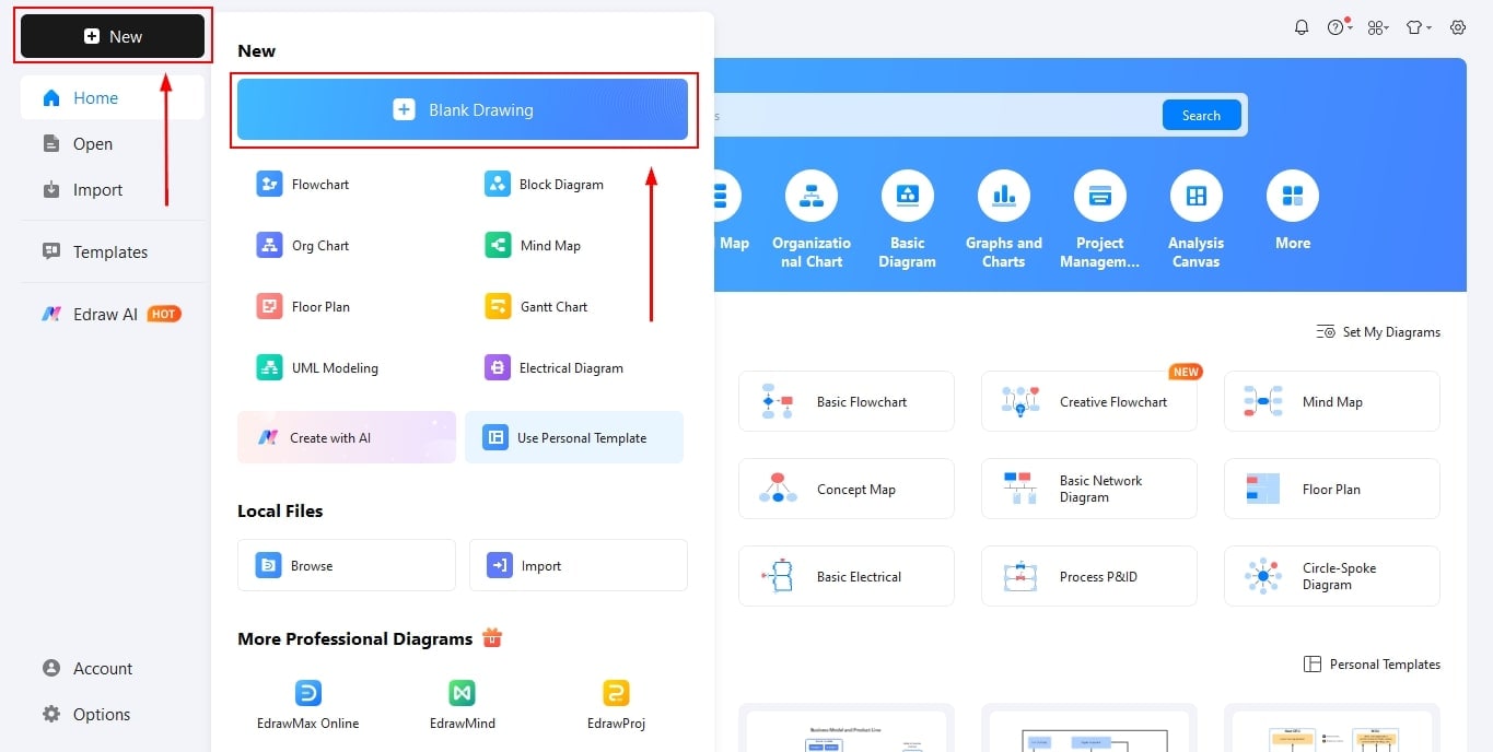
Step1 Open the Tool and Get Started
- Launch your preferred diagram-making software such as EdrawMax and select “New Diagram” from the dashboard.
- Choose a blank canvas or a biology-related template to ensure proper spacing and layout for cellular components.
- Set the page size and orientation (landscape works best) so all organelles of the endomembrane system fit clearly.
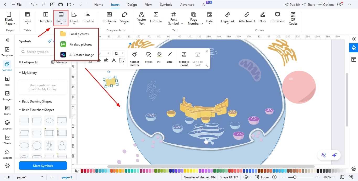
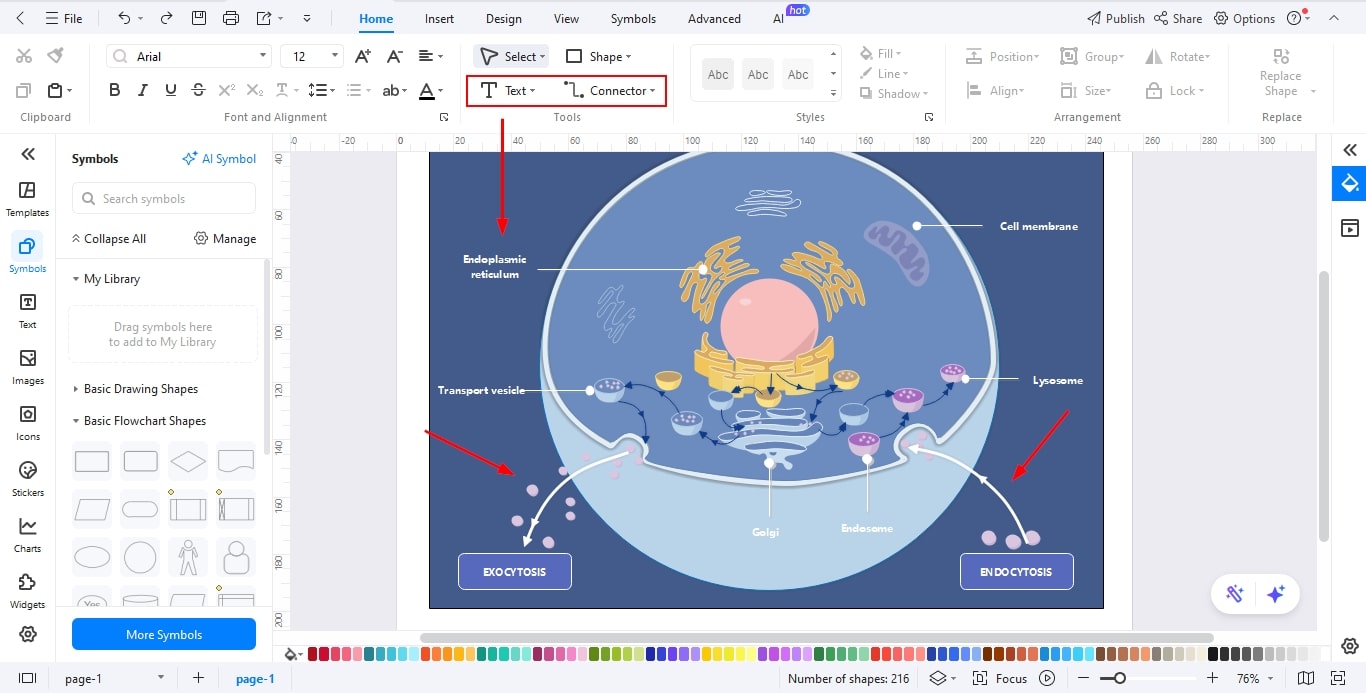
Step2 Add Elements and Pictures on the Canvas
- Use the symbol library or search bar to insert key endomembrane system components such as the nucleus, endoplasmic reticulum (rough and smooth), Golgi apparatus, vesicles, lysosomes, and plasma membrane.
- Drag and drop high-quality images or vector shapes onto the canvas to maintain a clean and professional look.
- Ensure each element is visually distinct to help viewers easily identify different organelles.
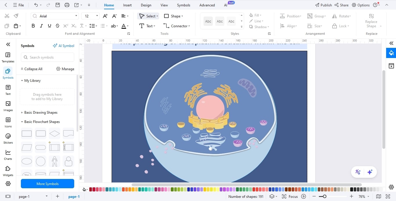
Step3 Rearrange the Images
- Arrange the organelles in a logical flow that represents the movement of proteins and lipids within the cell.
- Place the nucleus near the center, followed by the endoplasmic reticulum, Golgi apparatus, and vesicles in sequence.
- Adjust size, alignment, and spacing so the diagram looks balanced and avoids clutter.
Step4 Add Text and Label the Important Parts
- Add clear text labels for each organelle using readable fonts and consistent formatting.
- Include short descriptions explaining the function of each part, such as protein synthesis or transport.
- Use arrows or connector lines to show the direction of material movement within the endomembrane system.
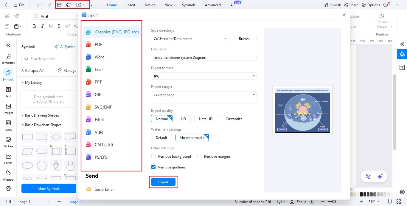
Step5 Export the Diagram
- Review the diagram carefully to ensure all components are correctly labeled and properly aligned.
- Select the Export option and choose a suitable format such as PNG, PDF, or JPEG based on your needs.
- Save the final diagram for use in presentations, assignments, or study materials.
Tip: If you want to increase the visual appeal of the diagram, you can choose to export it in the format of GIF, like this one:
Conclusion
In conclusion, the endomembrane system is not only crucial for the proper functioning of a cell but also for its survival. A detailed and properly labeled diagram makes it easier to understand such a complex system. EdrawMax is one of the best diagram tools available online that can be used to draw such diagrams. So, make sure to try it for your next biology diagram.
AI Diagram Generator
Enter your prompt. Upload files if needed. Generate diagrams, charts, or slides instantly.