


In living systems, proteins are vital biological molecules that perform a wide range of functions. It is easier to understand how things operate when one is aware of their structure. The purpose of this guide is to understand the four levels of protein structure and how to make their diagram.
Understanding protein structure is crucial for understanding the biological processes that occur in our bodies. By graphically relating structure to function, protein structure diagrams make biological concepts easier to understand. EdrawMax is an excellent tool to make such diagrams.
In this article
What Are the 4 Levels?
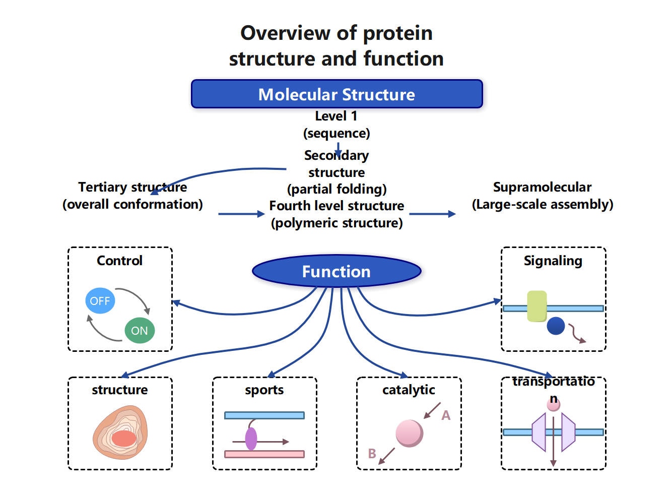
Proteins are classified into four structural levels; complexity increases with each level. A simple amino acid chain folds and forms a functional biological molecule. Here are four structures explained:
- Primary structure: It is simply a linear amino acid chain linked by peptide bonds. This chain also has information about proper folding.
- Secondary structure: It is formed when the chain folds into repeating patterns. For instance, alpha helices and beta sheets are stabilized with hydrogen bonds.
- Tertiary structure: It represents the three-dimensional structure of a single protein chain.
- Quaternary structure: It forms when multiple folded chains assemble to form one complex structure.
Structure–Function Relationship
The structure of a protein at every level determines its function. Primary structure determines how a protein will be folded as amino acids interact in a certain way. These interactions decide how the secondary and tertiary structure will be formed. Additionally, the 3-dimensional structure of the protein is also determined with the help of these interactions.
The final shape of protein creates binding pockets, active sites, and interaction surfaces. It allows them to perform specific functions, for instance, catalyzing and transporting. Small structural changes can reduce the efficiency of the protein.
Misfolded protein may lose its function partially or completely. Therefore, the structure-function relationship is crucial to explain normal protein activity.
Primary and Secondary Structure
All higher organizations are built upon the first two levels of protein structure. At this point, diagrams concentrate on straightforward folding patterns and linear sequences.
Amino Acid Sequence
The unique arrangement of amino acids connected by peptide bonds makes up the main structure. Genetic information determines the precise arrangement of each protein. Protein behavior can be changed by a single amino acid substitution. This chain also has information regarding how a protein will fold to be functional.
The main structure is frequently depicted in diagrams as:
- An uninterrupted series of linked amino acids
- Amino acid names or symbols that are abbreviated
- From the N-terminus to the C-terminus
These diagrams aid students in seeing how straightforward sequences develop into intricate systems.
Alpha Helices and Beta Sheets
The secondary structure is formed when an amino acid chain folds due to hydrogen bonding. The two most popular patterns are:
- Alpha helices: spiral-shaped coils
- Beta sheets: consist of folded, zigzag-like threads.
Secondary structure diagrams generally illustrate these shapes with ribbons or arrows. This stage demonstrates how regular folding patterns enhance the stability and shape of the protein before full 3D folding takes place.
How to Make a Protein Structure Diagram with EdrawMax
Protein structure diagrams need to be precise, logical, and easy to understand. EdrawMax is an easy-to-use diagramming tool. It provides specific biology and biochemistry materials, making it appropriate for everyone. Its built-in templates and symbols allow for the clear and visually appealing illustration of protein structures.
Step1 Choosing Biochemistry Templates
EdrawMax offers a number of ready-made templates for biochemistry that are excellent places to start.
- Standard symbols for protein chains, alpha helices, beta sheets, and amino acids are included in templates.
- Scientific conventions are followed when designing layouts.
- Using templates increases accuracy and cuts down on design time.
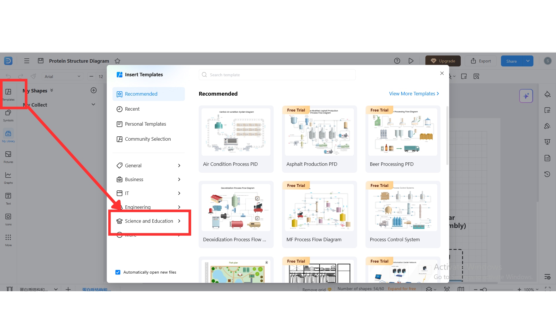
You need to choose the right template to get started. Tap on the “Template” button on the left panel, and from there choose “Science and Education,” and you can search for the desired biochemistry template.
Step2 Drawing Each Structure Level
Start by sketching the main structure as a chain of linear amino acids. Simple connected shapes or named symbols can be used for this.
- Show the N-terminus and C-terminus clearly.
- Labels should be used to show amino acid sequences.
- For ease of comprehension, keep the layout simple.
The secondary structure components, such as sheets and helices, should then be included in different parts of the diagram.
Step3 Illustrating Folding Patterns
EdrawMax permits the use of ribbon-style forms and curved connectors to depict tertiary structure.
- Demonstrate how the polypeptide chain folds into a small, three-dimensional shape.
- To show the direction of folding, use curves and arrows.
- Use color coding to identify different structural areas.
This stage makes it easier to see how folding produces useful protein structures.
Step4 Creating Multi-Level Diagrams
To display all four levels in a single picture, multi-level diagrams are helpful.
- Sort structures according to their complexity.
- On one side, arrange the primary and secondary structures.
- On the other side, show the tertiary and quaternary structures.
- Conceptual flow and comparison are enhanced by this arrangement.
Step5 Adding Chemical Bond Representations
Protein structure is stabilized by chemical interactions, which ought to be evident.
- Include symbols for disulfide bridges and hydrogen bonds.
- Identify hydrophobic and ionic interactions.
- Explain bond types using legends.
Adding these details is simple and doesn't overcrowd the graphic, thanks to EdrawMax's annotation tools. The finished diagram can be exported for use in reports, presentations, or instructional blogs.
Tertiary and Quaternary Structure
The tertiary and quaternary levels of protein structure represent their functional forms. Proteins at these levels interact with other molecules and carry out specific biological functions.
3D Folding
The full three-dimensional folding of a single polypeptide chain is referred to as the tertiary structure. Protein stability and activity depend on this folding. It is fueled by interactions between amino acid side chains.
- Maintained via disulfide bridges, hydrophobic contacts, ionic interactions, and hydrogen bonds
- Hydrophilic amino acids stay exposed while hydrophobic amino acids fold toward the inside.
- Each protein takes on a compact, distinct shape.
- Active sites and binding pockets essential for biological function are formed.
The way a protein interacts with ligands, substrates, or other proteins is determined by its tertiary structure. To properly arrange substrates and effectively catalyze reactions, enzymes rely on accurate folding. Protein function may be diminished or eliminated if tertiary folding is interfered with. Mutations or environmental elements like pH and temperature may affect the folding.
Multi-Subunit Complexes
Proteins made up of two or more polypeptide chains, referred to as subunits, have a quaternary structure. Each subunit contributes to the overall function of the protein complex and has a unique tertiary structure.
- Noncovalent interactions, like hydrogen bonds and ionic forces, are how subunits come together.
- Certain proteins also use disulfide bonds to fortify subunit connections.
- Enables cooperative behavior between subunits and enhances protein stability and functional control.
Hemoglobin is a well-known example of how many subunits cooperate to carry oxygen. Quaternary structure diagrams show various subunits in contrasting hues. It makes their arrangement and interactions easy to visualize and understand.
Explore More Free Biology Diagram Templates
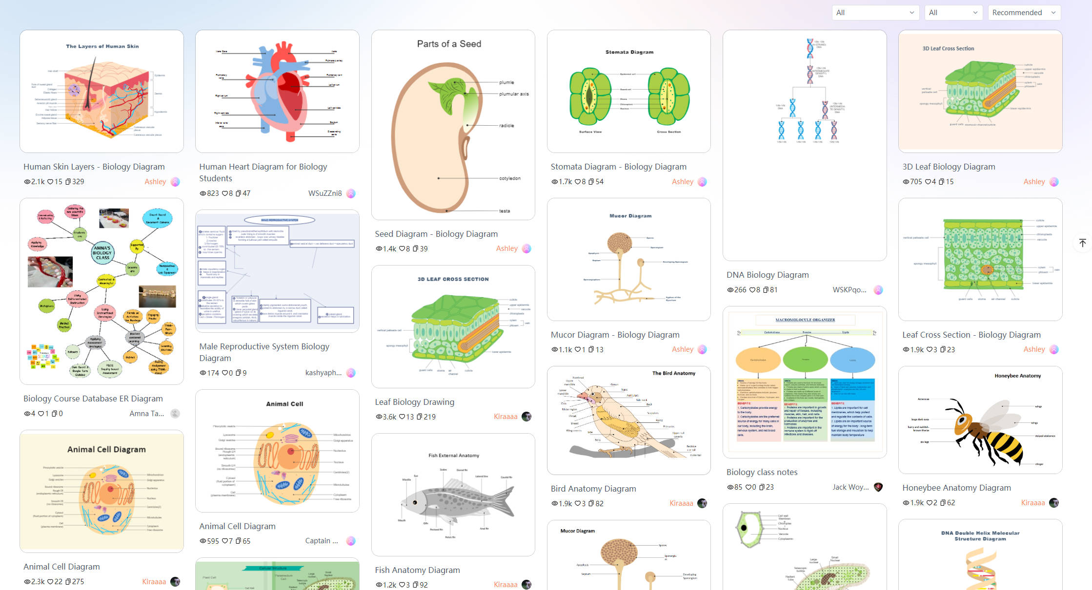
For researchers, teachers, and students who require accurate and comprehensible visual representations of protein arrangement, free protein structure templates are useful tools. These templates facilitate the process of creating diagrams. They provide pre-made layouts that follow recognized biological principles. You can easily find free templates on EdrawMax for any Biology diagram.
Conclusion
Understanding protein structure is crucial to learn about the biological processes happening in our bodies. By graphically relating structure to function, protein structure diagrams make biological concepts easier to understand. EdrawMax is an excellent tool to make such diagrams.
AI Diagram Generator
Enter your prompt. Upload files if needed. Generate diagrams, charts, or slides instantly.