


This guide describes two structurally and functionally different components of the endoplasmic reticulum, smooth and rough endoplasmic reticulum.
The endoplasmic reticulum is one of the complex organelles associated with protein and lipid manufacturing and transfer. The purpose of this blog is to help you understand how the Endoplasmic Reticulum works and how you can depict its structure and function in the form of a diagram.
Diagrams are a fundamental tool in biology education, helping students visualize complex cellular structures and processes. At the microscopic level, accurate illustrations clarify how organelles interact to sustain life.
One such organelle—the endoplasmic reticulum (ER)—plays a central role in protein and lipid production, making an endoplasmic reticulum diagram essential for understanding cell function.
This guide explains the structure and function of the ER, explores the protein transport pathway, and shows you how to create a clear and professional ER diagram.
In this article
What Is the Endoplasmic Reticulum?
The endoplasmic reticulum is a vital organelle found in almost all eukaryotic cells. Located in the cytoplasm, it forms an extensive, continuous membrane network that serves as the cell’s production and transport system.
The ER is involved in:
- Protein synthesis, folding, and modification
- Lipid and steroid synthesis
- Intracellular transport
- Calcium storage and detoxification
Structurally and functionally, the ER consists of two distinct regions:
- Rough Endoplasmic Reticulum (RER)
- Smooth Endoplasmic Reticulum (SER)
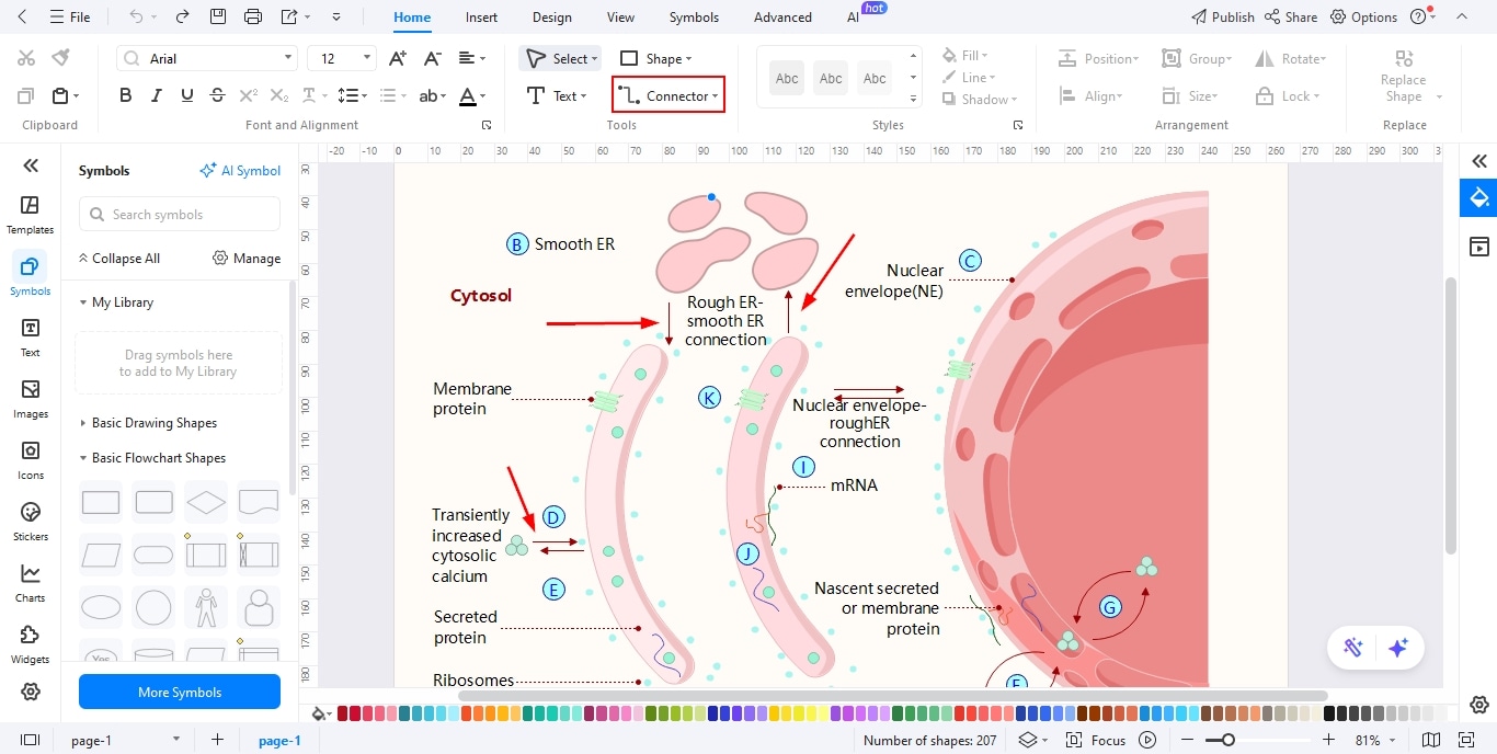
Rough Endoplasmic Reticulum
The rough ER is named for its ribosome-studded surface, which gives it a rough appearance under an electron microscope. Ribosomes are the molecular machines responsible for protein synthesis.
Key Characteristics
- Flattened membrane sacs called cisternae
- Located near the nucleus
- Abundant in cells that secrete large quantities of proteins (e.g., pancreatic and liver cells)
Protein Synthesis in Rough ER
- Ribosomes synthesize proteins destined for secretion, membranes, or lysosomes
- Newly formed polypeptides enter the RER lumen
- Chaperone proteins assist with proper folding
- Misfolded proteins are identified and degraded
- Correctly folded proteins are packaged for transport
Smooth Endoplasmic Reticulum
Unlike RER, the smooth ER lacks ribosomes and has a tubular appearance. It connects the rough ER to the peripheral cytoplasm.
The functions of smooth ER differ depending on the cell type. Generally, SER plays a role in lipid synthesis, steroid hormone production, detoxification, carbohydrate metabolism, calcium storage, and regulation.
ER Structure and Cellular Organization
The endoplasmic reticulum accounts for more than 50% of the total membrane system in an animal cell. It is composed of interconnected cisternae and tubules, forming a continuous lumen known as the cisternal space.
The ER membrane is continuous with the outer nuclear envelope, explaining its close functional relationship with the nucleus. Genetic instructions stored in the nucleus are translated and executed within the ER.
Protein Synthesis in Rough ER
Rough ER is basically responsible for protein handling from synthesis to modification to quality control and transport.
- Ribosomes synthesize proteins that are meant to be secreted from the cell or within the cell.
- After synthesis, proteins are transferred to the RER lumen, where they undergo proper folding with the help of chaperone proteins.
- RER has an excellent quality control system that ensures the transportation of properly folded proteins, while misfolded proteins are subjected to degradation.
Lipid Synthesis in Smooth ER
Smooth endoplasmic reticulum is a vital site for various lipid formation, including cholesterol and phospholipids. These are fundamental building blocks of all kinds of cellular membranes. Cells in which there is high production of steroid hormones contain abundant SER, for instance, the ovaries and testes.
Calcium Storage
The smooth ER—and its muscle-specific form, the sarcoplasmic reticulum (SR)—plays a crucial role in calcium homeostasis.
- Stores large quantities of calcium ions (Ca²⁺)
- Releases calcium for signaling, muscle contraction, and heart rhythm
- Rapidly pumps calcium back to prevent cytotoxicity
Disruption in calcium regulation is linked to muscle disorders, neurodegenerative diseases, and apoptosis.
Protein transport pathway
The protein transport pathway ensures that newly synthesized proteins reach their correct destinations inside or outside the cell. Most secretory, membrane, and lysosomal proteins begin this journey in the ER.
Proteins that pass quality control are packaged into vesicles and transported along the cytoskeleton toward the Golgi apparatus.
ER to Golgi Transport
The ER-to-Golgi protein transport pathway is the secretory pathway's first step, where correctly folded proteins exit the ER and enter the Golgi complex. Here are the key steps:
- Cargo Selection and Budding (COPII)
- Occurs at ER exit sites (ERES)
- COPII coat proteins (Sar1, Sec23/24, Sec13/31) form vesicles
- Cargo proteins are selectively packaged
- Vesicular Tubular Clusters (VTCs)
- COPII vesicles fuse to form VTCs
- Serve as an intermediate compartment between the ER and the Golgi
- Fusion with cis-Golgi
- VTCs fuse with the cis face of the Golgi
- SNARE proteins ensure correct membrane fusion
- Retrograde Transport (COPI)
- COPI vesicles return ER-resident proteins
- Maintains protein balance and quality control
- Movement Through the Golgi
- Proteins undergo modifications (e.g., glycosylation)
- Sorted at the trans-Golgi network
- Delivered to lysosomes, the plasma membrane, or secreted
How to Make an Endoplasmic Reticulum Diagram with EdrawMax
Creating a detailed endoplasmic reticulum diagram is simple with EdrawMax, a user-friendly diagramming tool designed for both beginners and professionals.
Why Use EdrawMax?
- Drag-and-drop biological symbols
- Pre-designed ER, ribosome, vesicle, and Golgi shapes
- Auto-alignment and connector tools
- High-resolution export options
How to Create an Endoplasmic Reticulum Diagram on EdrawMax
Below is a step-by-step, bullet-point guide on how to create an Endoplasmic Reticulum (ER) diagram using EdrawMax. Follow these steps to complete you ER diagram.
Step1 Open the Tool and Get Started
- Launch EdrawMax on your computer or access the online version through your browser and sign in.
- From the home dashboard, click on “New” and choose a blank canvas or search for a cell biology diagram template.
- Adjust the page layout, orientation, and canvas size to provide enough space for labeling both Rough ER and Smooth ER clearly.

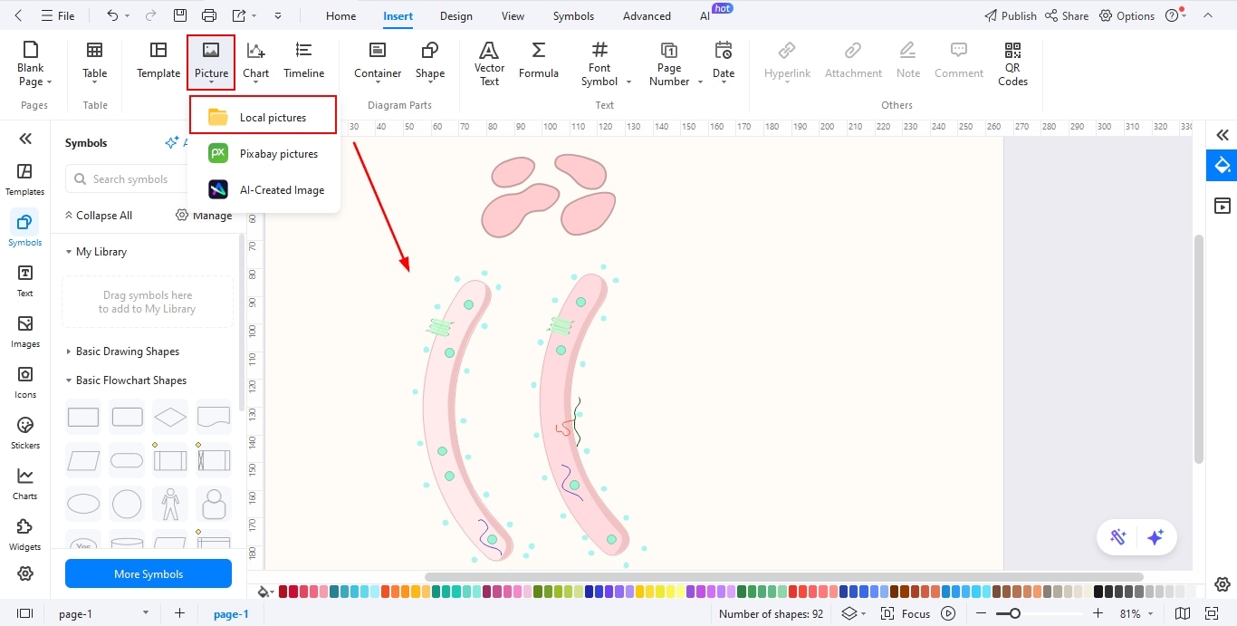
Step2 Insert Images
- Open the Symbols Library and browse the biology or cell structure categories available in EdrawMax.
- Insert shapes or icons representing the Endoplasmic Reticulum, ribosomes, and nucleus for better context.
- You can also upload high-quality ER images from your device to enhance scientific accuracy and visual appeal.
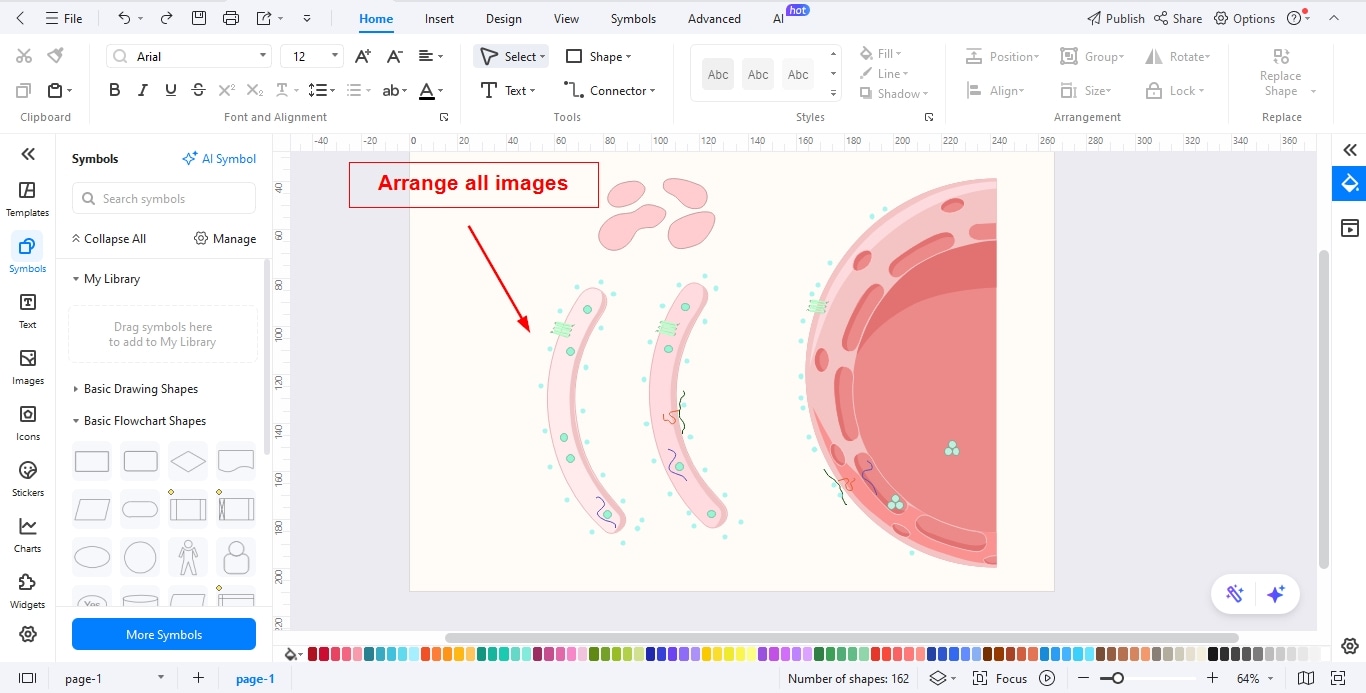
Step3 Arrange the Images
- Position the ER structure around the nucleus to reflect its actual location inside the cell.
- Separate and clearly distinguish Rough ER (with ribosomes) from Smooth ER (without ribosomes).
- Use alignment and spacing tools to maintain a neat layout and avoid overlapping elements.
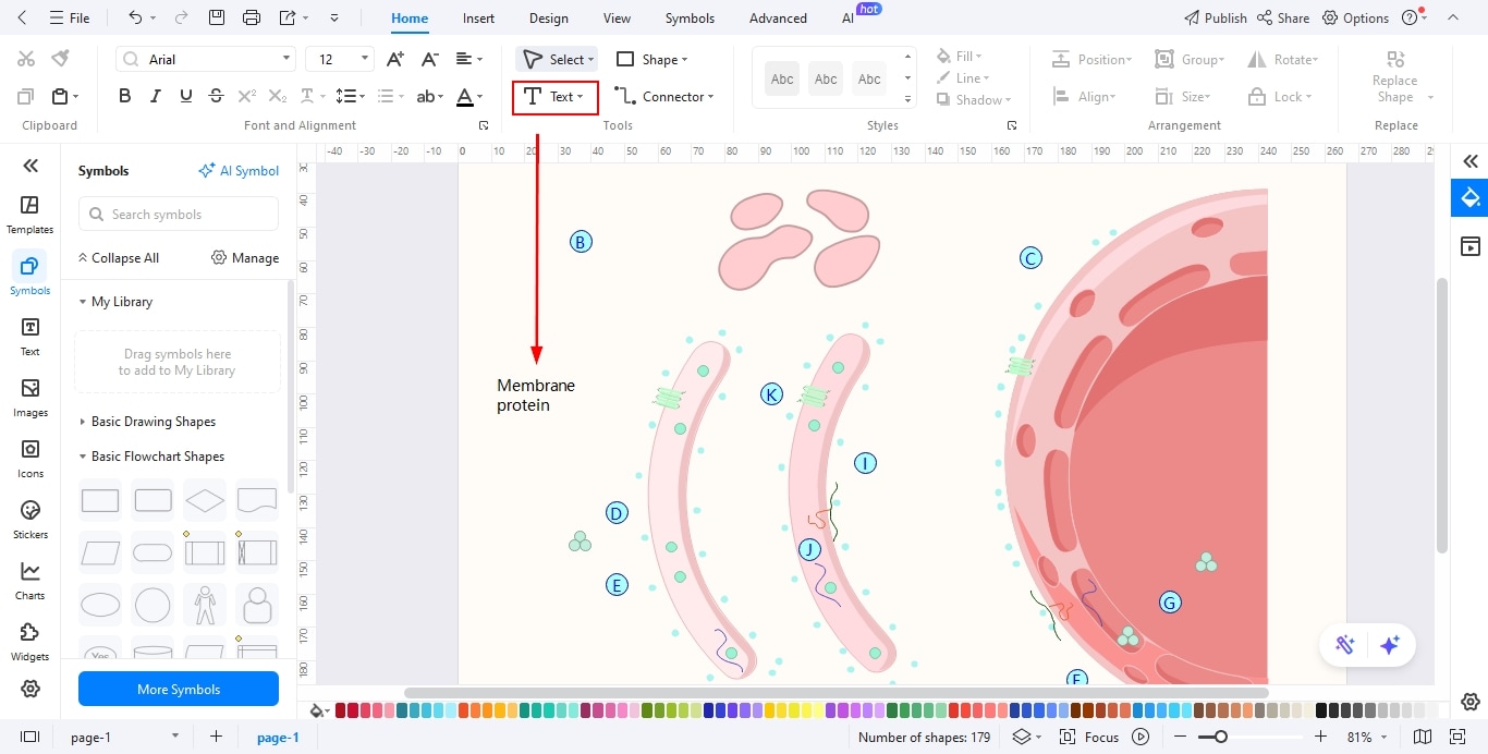
Step4 Add Text for Each Phase and Important Notes
- Add text boxes to label important components such as cisternae, ribosomes, lumen, and membranes.
- Write brief explanations for Rough ER and Smooth ER functions, including protein synthesis, lipid production, and detoxification.
- Include important notes to highlight key differences, such as the presence or absence of ribosomes and their specific cellular roles.
Step5 Add Relationships Among Different Phases
- Use the Connector Tool to link labels and notes to their corresponding parts of the ER.
- Add arrows to show the flow of proteins from the Rough ER to other organelles like the Golgi apparatus.
- Apply different line styles or colors to visually separate functional relationships within the diagram.
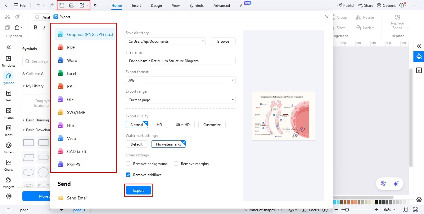
Step6 Export Your Design
- Carefully review the diagram for clarity, accuracy, and proper alignment.
- Click File and find the Export options and find the desired format, such as PNG, PDF, PPT, or Word.
- Save or share your final Endoplasmic Reticulum diagram for use in presentations, exams, or study materials.
In the end, this is what you will get:

Conclusion
Understanding the endoplasmic reticulum is essential for grasping how cells synthesize, modify, and transport vital molecules. A well-labeled endoplasmic reticulum diagram simplifies these complex processes, while tools like EdrawMax make diagram creation fast and professional.
With intuitive templates and powerful customization features, anyone—from students to educators—can create clear, accurate ER diagrams and confidently explain the protein transport pathway.
AI Diagram Generator
Enter your prompt. Upload files if needed. Generate diagrams, charts, or slides instantly.