


The animal cell is the fundamental unit of life, a complex, membrane-bound structure housing vital organelles like the nucleus and mitochondria. Understanding its intricate structure and functions is essential for appreciating human biology, health, and the diversity of life.
This guide uses a detailed animal cell diagram to walk you through its key components and their crucial roles.
In this article
- What Is an Animal Cell?
- Animal Cell vs Plant Cell: Key Structural Differences
- Why Understanding Animal Cell Biology Is Essential
- 11 Essential Cell Organelles and Their Functions
- Understanding Cell Organelle Functions in Detail
- How to Draw an Animal Cell Diagram Step-by-Step
- How to Create the animal Cell diagram on EdrawMax
- Final Thoughts
What Is an Animal Cell?
The animal cell is a structural and functional part of the animal body. The bodies of animals may either be unicellular or multicellular, and the cells are membrane-bound with various organelles. Animal cells are of different shapes and sizes - the largest being an ostrich egg, which has a 5-inch diameter, and the smallest being a neuron, which has a diameter of 100 microns. Being eukaryotic cells, they contain organelles that are enclosed by a membrane.
Every cell is surrounded by a plasma membrane that isolates its internal environment from the external environment. The protoplasm is made up of the cytoplasm and the nucleus, which are found in the cell. The cytoplasm comprises the cytosol and a number of organelles. The cell membrane acts as a selective barrier that ensures cell integrity.
Animal Cell vs Plant Cell: Key Structural Differences
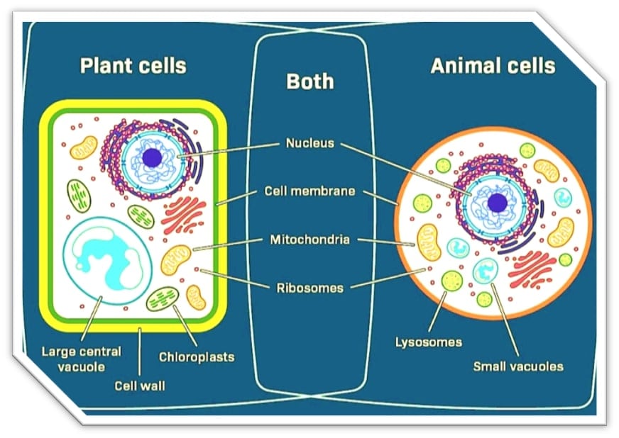
The animal and plant cells are eukaryotic and contain membrane-bound organelles. Nevertheless, these two classes of cells do have some major differences. The plant cells are rigid with cell walls and cell membranes, whereas the animal cells have a single cell membrane.
The mitochondria can be found in both animal and plant cells, but chloroplasts are found in plant cells. Besides, plant cells possess large vacuoles, whereas animal cells possess numerous small vacuoles.
The cell wall of the plant cells contains cellulose. There are no walls to animal cells. Plant cells do not tend to have centrioles and cilia, whereas animal cells possess all of these parts.
Why Understanding Animal Cell Biology Is Essential
The animal cells have various functions in the human body. Their organelles and structure are essential for such functions of the body. Biology is a practical subject that needs visual representation and images in order to comprehend the structure and functions of those mechanisms. Biology diagrams come in several types, which make teaching and learning more convenient.
Cells form the structural and functional components of the body, and, therefore, to know the structure and functions of an animal body, one must study animal cells. It has several organelles and nuclei, and it complicates the learning process. The animal cell diagram may also be difficult to explain on paper; therefore, students should focus on the essential organelles for a better understanding of cell biology.
11 Essential Cell Organelles and Their Functions
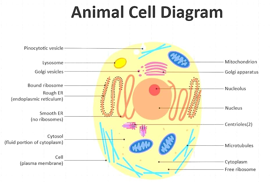
Based on the diagram of animal cells, the organelles of an animal cell are the cell membrane, cytosol, cytoskeleton, nucleus, ribosomes, endoplasmic membrane, and vesicles, among others. Further discussion of each organelle is given below;
Nucleus: The Cell Control Center and Cellular Regulation of the Genetic Material
The nucleus is the location where DNA is kept and regulates the cellular functions, such as growth, metabolism, and reproduction. It is enclosed by a nuclear envelope and has the nucleolus, which houses the ribosomes.
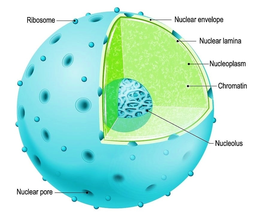
The largest organelle in a eukaryotic cell is the nucleus, which is believed to be the control center of the cell. It holds a majority of the DNA of the cell, which constitutes chromosomes and carries the genetic code for producing proteins. The nucleus has the role of controlling gene expression, such as the production of proteins that are made by the cell.
Mitochondria: The Powerhouse of the Cell
Mitochondria are the energy factory of the cell. They generate cellular respiration, which is essential to the organs and muscles.
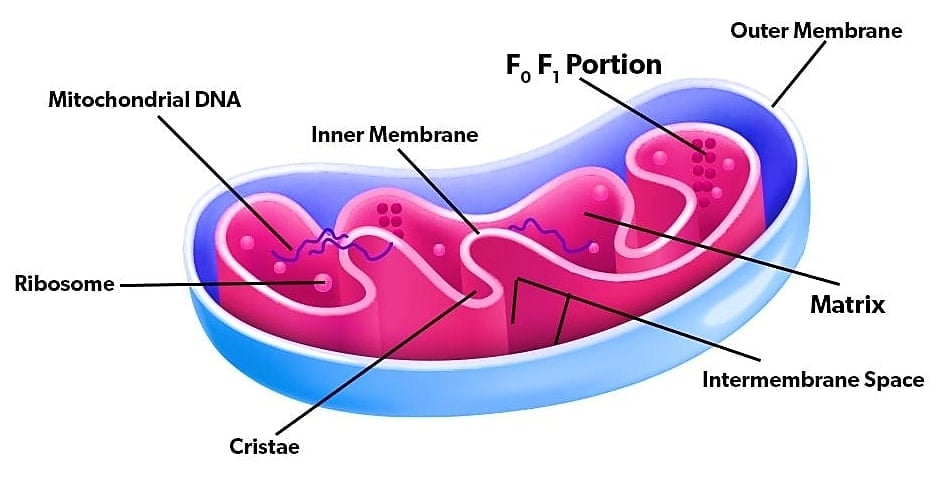
Why Are Mitochondria Essential for Cellular Function?
In animal cells, mitochondria are referred to as the powerhouse of the cell. They produce energy by breaking down glucose and oxygen into ATP (adenosine triphosphate) by means of cell respiration. The main energy currency is ATP, used in different cellular processes. Cell signaling and apoptosis (programmed cell death) are also facilitated by the mitochondria. This organelle makes sure that animal cells are supplied with the energy required to operate.
Endoplasmic Reticulum (ER): Manufacturing and Transport System
The endoplasmic reticulum (ER) is a series of continuous membranes that creates a net of tubules and sacs all over the cytoplasm. There are two types: Rough ER and Smooth ER.
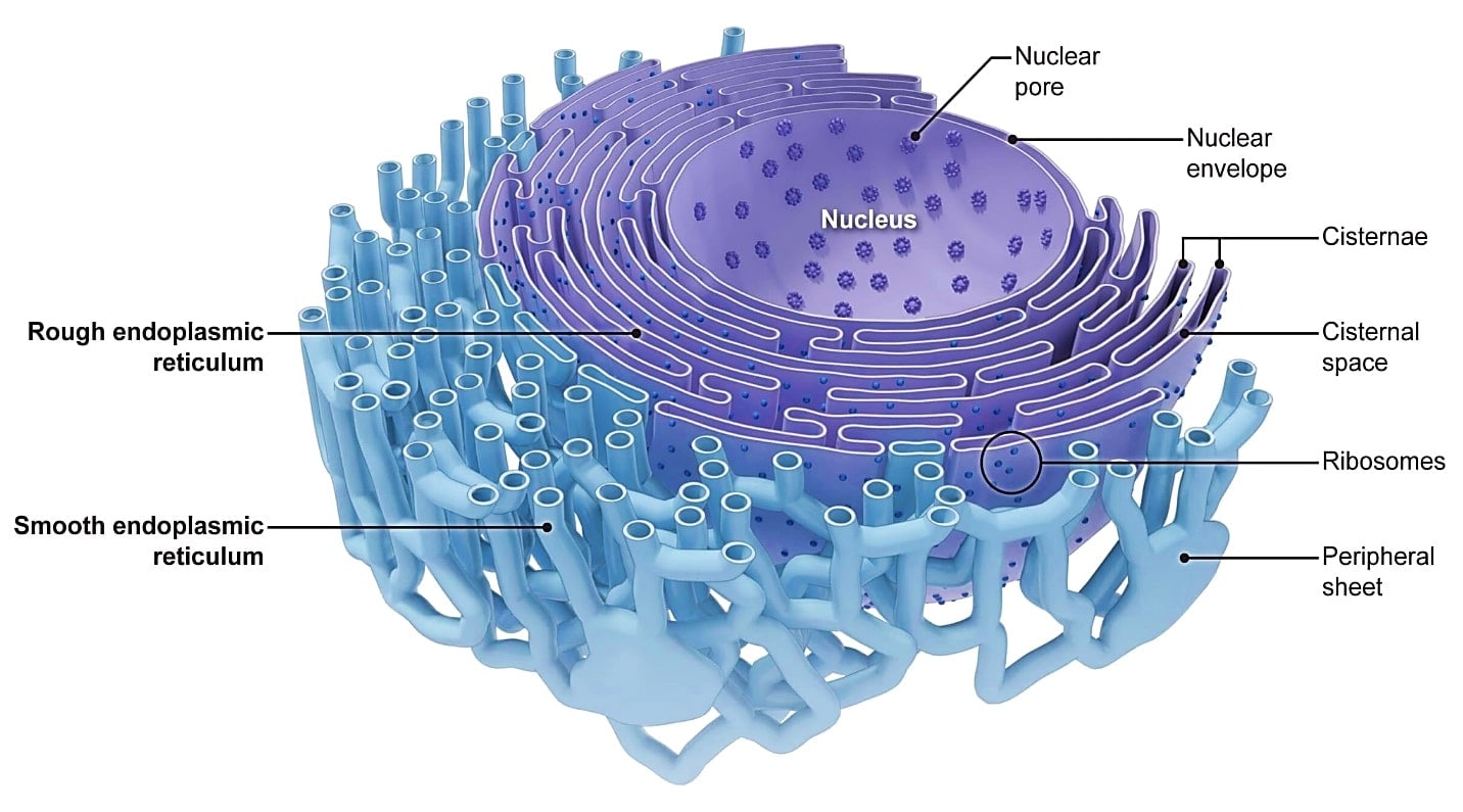
Rough Endoplasmic Reticulum (RER): Protein Factory
Rough endoplasmic reticulum (RER) is an important part of protein production. The notion that the RER is the ribosomal attachments on the surface resembles the smooth ER (SER), which lacks ribosomes. The RER can be morphologically differentiated because of the formation of a series of convoluted, membrane-flattened sheets called cisterna, which develop around the nucleus and stretch across the cytoplasm.
Smooth Endoplasmic Reticulum (SER): Lipid Processing and Detoxification
Smooth ER is an organelle in the cell that consists of a tube-shaped structure with multiple folds. Cells contain a specialized smooth ER that performs numerous functions. It produces steroids, lipids, and phospholipids, including those from the testes, ovaries, and sebaceous glands, which are rich in smooth ER. It also performs the natural metabolism of carbohydrates and steroids, and drug detoxification, through protein receptors on the cell membrane. It also involves regulating and controlling the level of calcium ions in muscle cells.
Golgi Apparatus: Packaging and Distribution Center
The Golgi apparatus is involved in the transportation, alteration, and packaging of proteins and lipids into vesicles for delivery to specific destinations. It is found in the cytoplasm adjacent to the endoplasmic reticulum and close to the cell nucleus.
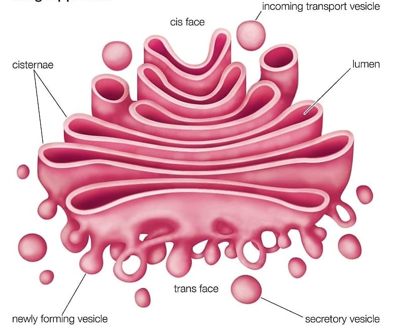
Golgi Apparatus: Processing and Shipping
The Golgi apparatus, Golgi complex, Golgi body, or Golgi is a cell organelle present in the majority of eukaryotic cells. It is a part of the endomembrane system within the cytoplasm, and it packages proteins into vesicles (membrane-bound) within the cell. These vesicles are transported to the specific destination of the other organelles. It sits between the secretory, lysosomal, and endocytic pathways. It is especially important to process secreted proteins, which have a set of glycosylation enzymes that attach different sugar monomers to proteins as the proteins pass through the apparatus.
The molecules are typically transported by transport vesicles to the rough ER to the cis face of the Golgi stacks, where they fuse with the Golgi membrane and are sorted according to their next destination. They are transported to the Golgi cisternae, where they are remodeled and undergo additional changes.
The resulting modified protein or lipid molecules then leave the trans face of the Golgi stacks into which they are secreted out of the cell, or into which they are transported into another cell compartment. The intricate system of membranes and vesicles of the Golgi stacks, where molecules are incorporated, is also referred to as the cis Golgi network.
Lysosomes, Ribosomes, and Other Key Organelles
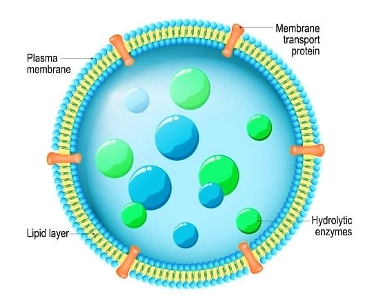
Lysosomes: The Digestive System
Lysosomes are the most common organelle found in animal cells. Lysosomes are very important for cellular recycling and contain enzymes to digest the undesirable materials, cellular waste, and cell debris. The Lysosomes bear a very low pH, and the enzymes contained within them are specifically adapted to operate under such conditions.
Lysosomes: Waste Disposal and Recycling
Lysosomes are circular organelles that are enclosed in a membrane and contain digestive enzymes that assist in digestion, excretion, and the cell renewal process.
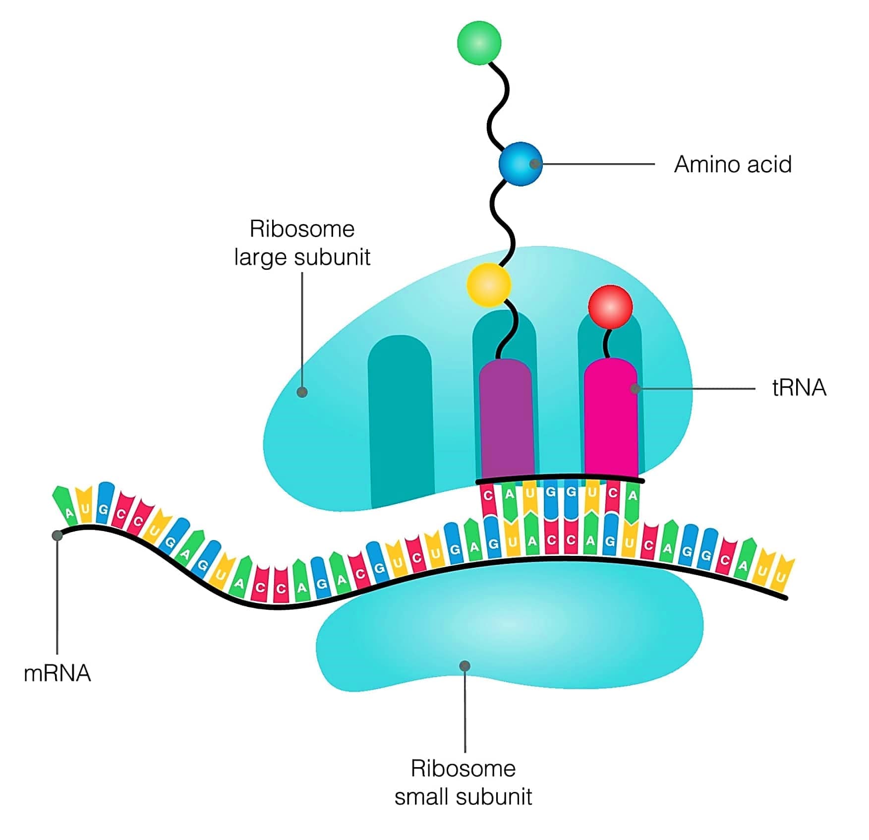
Ribosomes: Protein Synthesis Machines
Ribosomes are small bodies made up of RNA and proteins that synthesize proteins within the cell. They are either released freely in the cell or located with the endoplasmic reticulum. Ribosomes are attached to the rough ER surface, and the Smooth ER does not have any ribosomes. The Golgi body assists in the packaging and release of the ER-received proteins synthesized by the ribosomes.
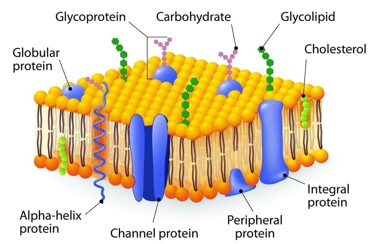
Cell Membrane: The Protective Barrier
The major function of the cell membrane is to protect the cell. It regulates the entry of nutrients and other small particles into the cell. Owing to this fact, cell membranes are regarded as semi-permeable or selectively permeable membranes. Each animal cell is enclosed by the plasma membrane, which determines the integrity of the structure and regulates the flow of materials in and out of the cell. One of the characteristics of the animal cell structure is the lack of a rigid cell wall.
How Does Cell Membrane Selective Permeability Work?
What is the mechanism of Cell Membrane Selective Permeability? The eukaryotic cell is characterized by animal cells, which are surrounded by a plasma membrane and have a nucleus, which is membrane-enclosed, and organelles. Animal cells lack a cell wall as opposed to the eukaryotic cells of plants and fungi. This property was lost in the distant past among single-celled organisms that gave rise to the kingdom Animalia.
Understanding Cell Organelle Functions in Detail
Cell Membrane: Selective Permeability and Transport
The cells are bounded by a thin plasma membrane, which isolates the internal contents of each cell against the external environment. The cell membrane acts as a selective barrier that keeps the cells intact.
The cell membrane is commonly referred to as the plasma membrane, and there are a few important functions that it performs:
- Regulates cellular inflow and outflow.
- Offers security and order.
- Possesses cell-to-cell-communication receptors.
- Helps keep the cells at homeostasis.
- Anchors the cytoskeleton
Cytoplasm: The Cell's Working Environment
A multicellular organism has its internal cytoplasmic compartment in all the living cells and has a nucleus located in the cytoplasm. The jelly-like substance of the cell, called Cytosol, furnishes the fluid medium by which the biochemical reactions occur.
The cell is filled with a gooey material known as the cytoplasm that offers the medium through which chemical reactions can take place. The control center of the cell is known as the nucleus, which harbors the genetic material and plays an important role in the cellular functions, including growth, reproduction, and metabolism.
Cytoplasm is not a mere passive medium, but it is:
- Location of numerous metabolic reactions.
- A storage area for nutrients.
- A movement medium through which materials are transported through organelles.
- The place of protein synthesis (in ribosomes).
- The cytoskeleton supports the cell structure.
How to Draw an Animal Cell Diagram Step-by-Step
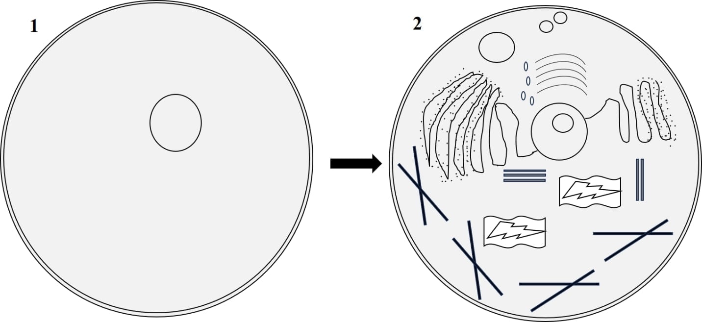
1) Start with the description of the cell cross-section.
Since it is a cross-section, a portion of the cell has been removed so you can look inside. Trace a major animal cell-shaped figure using a curved line. And then put in another shape of cell to the first. Lastly, sketch a parallel line to the bottom of the inner shape of the cell, connecting to the outer shape. Now you have your cell membrane, the three-dimensional edge, where you have peeled it off, and the cytoplasm, which is inside the cell.
2) Include organelles:
Add all the different organelles discussed above, including mitochondria, ribosomes, lysosomes, Golgi apparatus, and centrioles. Simple shapes can be used to depict every organelle.
3) Start labelling your cell structures and organelles.
What do you know the names of each? The large ovals are Lysosomes. There is the rough endoplasmic reticulum placed above and below the nucleus. The nuclear pores are represented by small ovals on the surface of the nucleus, and the core of the nucleus is the nucleolus. The structures are bean-shaped and are referred to as mitochondria, and the inter-structure is a fluid known as cytoplasm.
4) Finish labeling your cell.
The smooth endoplasmic reticulum and the Golgi apparatus are irregular structures containing lines within them. The centriole is an irregular shape that is without texture. Ribosomes are made up of groups of small ovals, and the outer margin of the figure is the cell membrane.
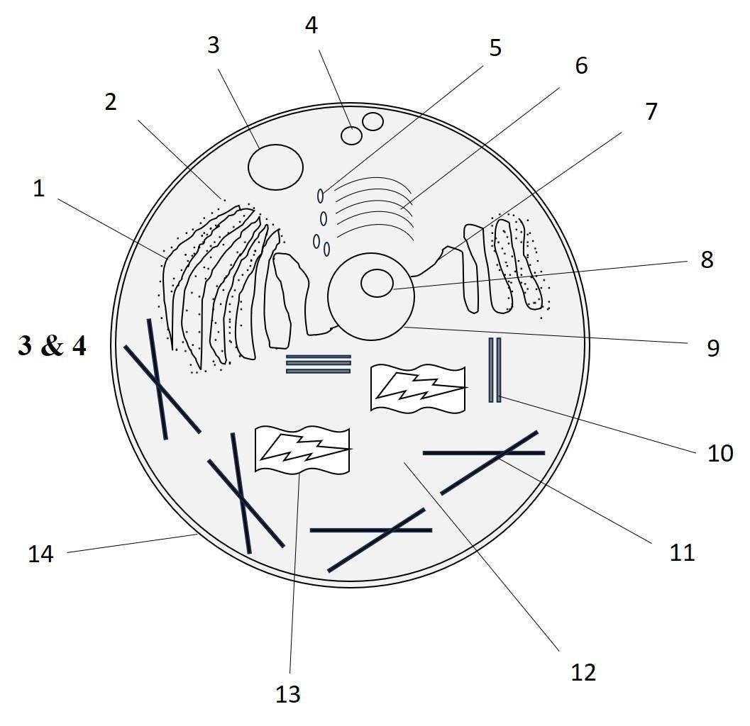
| Sr. Number | Cell Organelle Name | Sr. Number | Cell Organelle Name |
| 1 | Rough Endoplasmic Reticulum | 8 | Nucleolus |
| 2 | Ribosomes | 9 | Nucleus |
| 3 | Lysosomes | 10 | Centrioles |
| 4 | Pinocytotic Vesicle | 11 | Microtubules |
| 5 | Golgi Vesicle | 12 | Cytoplasm |
| 6 | Golgi Apparatus / Golgi Body | 13 | Mitochondria |
| 7 | Smooth Endoplasmic Reticulum | 14 | Plasma Membrane |
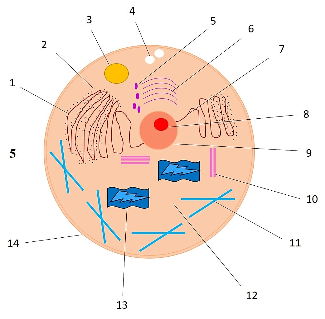
5) Fill in your drawing of the anatomy of an animal cell.
Although the colors in our drawing are not used to depict the original coloration, they contribute to the differentiation of the various parts.
Labeling Guidelines: What Organelles to Include
Stain the other part of the cell, which is referred to as cytosol or cytoplasm, with a liquid in where the organelles exist. Label it as you label it, because then you do not need to go back to the model and find each organelle of the cell. Draw the organelles in a checklist and cross them as you make them to ensure that you have not missed anything in your diagram.
Always remember to draw an animal cell and not a plant cell. To have an overall animal cell diagram, make sure to include and label all these important organelles:
- Cell membrane,
- Nucleus,
- Nucleolus,
- Mitochondria,
- Rough endoplasmic reticulum,
- Smooth endoplasmic reticulum,
- Golgi apparatus,
- Lysosomes,
- Ribosomes,
- Cytoplasm,
- Centrioles.
Color-Coding Tips for Better Cell Visualization
Each structure and its role are described on a numbered list. The students locate the cell parts using the numbers on a diagram and color them. It is easy, and students can accomplish this without a textbook or background knowledge.
Draw the cell on a piece of paper. Name each organelle in the diagram and indicate each organelle by a different color. Trace the cell membrane, and it will be the outline of the cell. Draw the cytoskeleton. To have successful color-coding of diagrams of animal cells, one should bear in mind the following:
- Neighboring organelles are to be used with contrasting colors.
- Important organelles such as the nucleus and mitochondria should be in bright colors.
- Related structures should be given similar color families (e.g., rough and smooth ER of darker and lighter blues).
- Prepare a color key at the bottom of your diagram.
- Draw many cells to compare them, and use the same color.
How to Create the animal Cell diagram on EdrawMax
EdrawMax offers ready-made symbols and smart design features that make biology diagrams easy to build. The step-by-step process below will guide you from setup to final export with ease.
Step1 Open the Tool and Get Started
- Launch EdrawMax on your computer and sign in to your account.
- Choose a blank canvas or an Animal Cell Diagram template to save time.

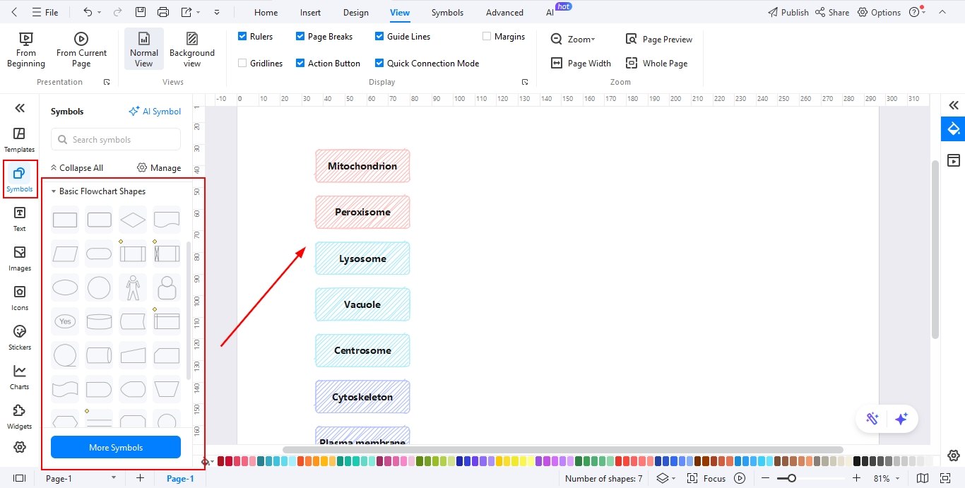
Step2 Drag Elements Needed
- Open the Symbol Library panel and enable the Biology and Cell Structure libraries.
- Drag essential animal cell components such as the nucleus, mitochondria, ribosomes, ER, Golgi apparatus, lysosome, and cell membrane onto the canvas.
- Resize and duplicate elements as needed to maintain accuracy and proportion.
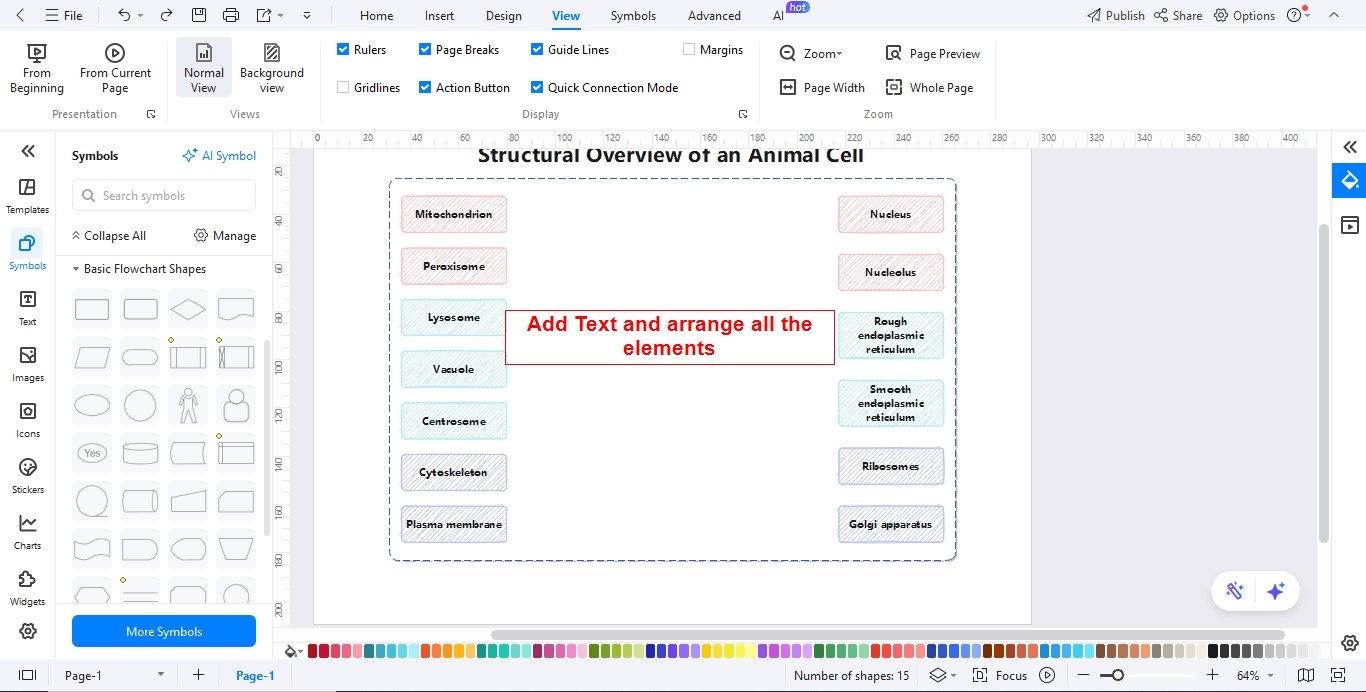
Step3 Arrange the Elements and Add Text
- Position each organelle correctly within the cell boundary.
- Use the Text tool to label each part clearly with readable fonts.
- Adjust font size, color, and alignment to keep the diagram neat and professional.
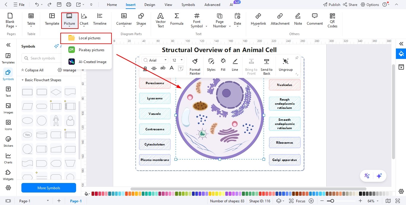
Step4 Insert Image from Your Computer
- Go to Insert > Image > Import to upload a reference image if needed.
- Place the image beside or behind the diagram for guidance.
- Adjust transparency or lock the image to avoid accidental movement.
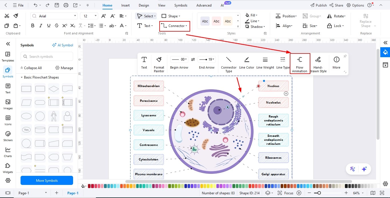
Step5 Add Relationships in the Form of Animations
- Use the Connector tool to show relationships between organelles.
- Apply arrows or curved connectors to indicate functional links.
- Add animations or dynamic effects (if presenting) to visually explain processes like protein synthesis or energy production.
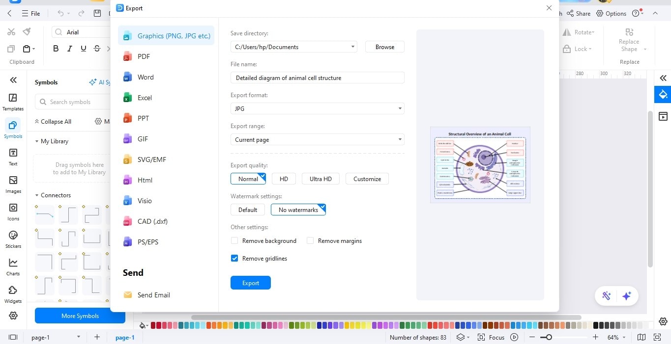
Step6 Export the File
- Click Export in the top menu.
- Choose your preferred format, such as PNG, JPG, PDF, Word, or PowerPoint.
- Set resolution and quality options, then save or share your final animal cell diagram.
Final Thoughts
To sum up, the study of animal cell structure is essential in the study of biology. With the help of the elaborate diagram templates and method of drawing on the diagrams described in this guide, students will be able to draw transparent and informative visualizations that will improve their understanding of cellular biology.
AI Diagram Generator
Enter your prompt. Upload files if needed. Generate diagrams, charts, or slides instantly.