


Meiosis is a two-stage cell division process that produces four genetically distinct haploid gametes. It is vital for sexual reproduction and driving essential genetic diversity.
This guide fully explains the 8 stages of Meiosis I and II, including crossing over. Learn to draw the process accurately using our step-by-step tutorial and free templates.
In this article
- What Is Meiosis? Understanding Sexual Reproduction
- Meiosis I vs Meiosis II: The Two-Stage Division Process
- Why Meiosis Creates Genetic Diversity in Genetics?
- The 8 Stages of Meiosis
- How Does Meiosis Differ from Mitosis?
- How to Draw a Meiosis Diagram: Step-by-Step Tutorial
- Bonus: Tips for Visualizing Crossing Over and Genetic Recombination
- FAQs About Meiosis
What Is Meiosis? Understanding Sexual Reproduction
Meiosis is a type of cell division that occurs in germ cells. It produces 4 genetically unique haploid cells from one diploid parent. It is essential for sexual reproduction because it produces gametes, e.g., eggs and sperm. Gametes further take part in sexual reproduction to produce a diploid offspring.
Meiosis I vs Meiosis II: The Two-Stage Division Process
The meiosis process completes in two stages, Meiosis I and Meiosis II, each with four distinct stages: Prophase, Metaphase, Anaphase, and Telophase.
In Meiosis I, also known as reductional division, the number of chromosomes is cut in half. During this process, homologous chromosomes got separated. Two daughter cells are formed with one chromosome from each pair. This stage brings genetic diversity in individuals
Meiosis II, also known as equational division, resembles the process of mitosis and produces 4 haploid daughter cells by separating the sister chromatids. The key differences between these two stages are explained in the table:
| Meiosis I | Meiosis II |
| Reduction in chromosomes from diploid to haploid | Further reduction does not occur, and the number of chromosomes remains the same |
| Synapsis occurs, and homologous chromosomes separate. | Synapsis does not occur, and sister chromatids separate. |
| Crossing over happens, and genetic material is exchanged | Crossing over does not happen |
| At the end of this stage, 2 haploid cells are produced | At the end of this stage, 4 haploid cells are produced |
Why Meiosis Creates Genetic Diversity in Genetics?
Meiosis creates genetic diversity that is essential for evolution in species by producing genetically unique haploid cells. The process of meiosis brings diversity in an individual’s genome by following key phases:
- Crossing over is when homologous chromosomes exchange their genetic material. It creates new combinations of genes in offspring.
- Independent assortment of chromosomes happens in the metaphase of Meiosis I. In this phase, the orientation of chromosomes is random. It means various genetic combinations can form.
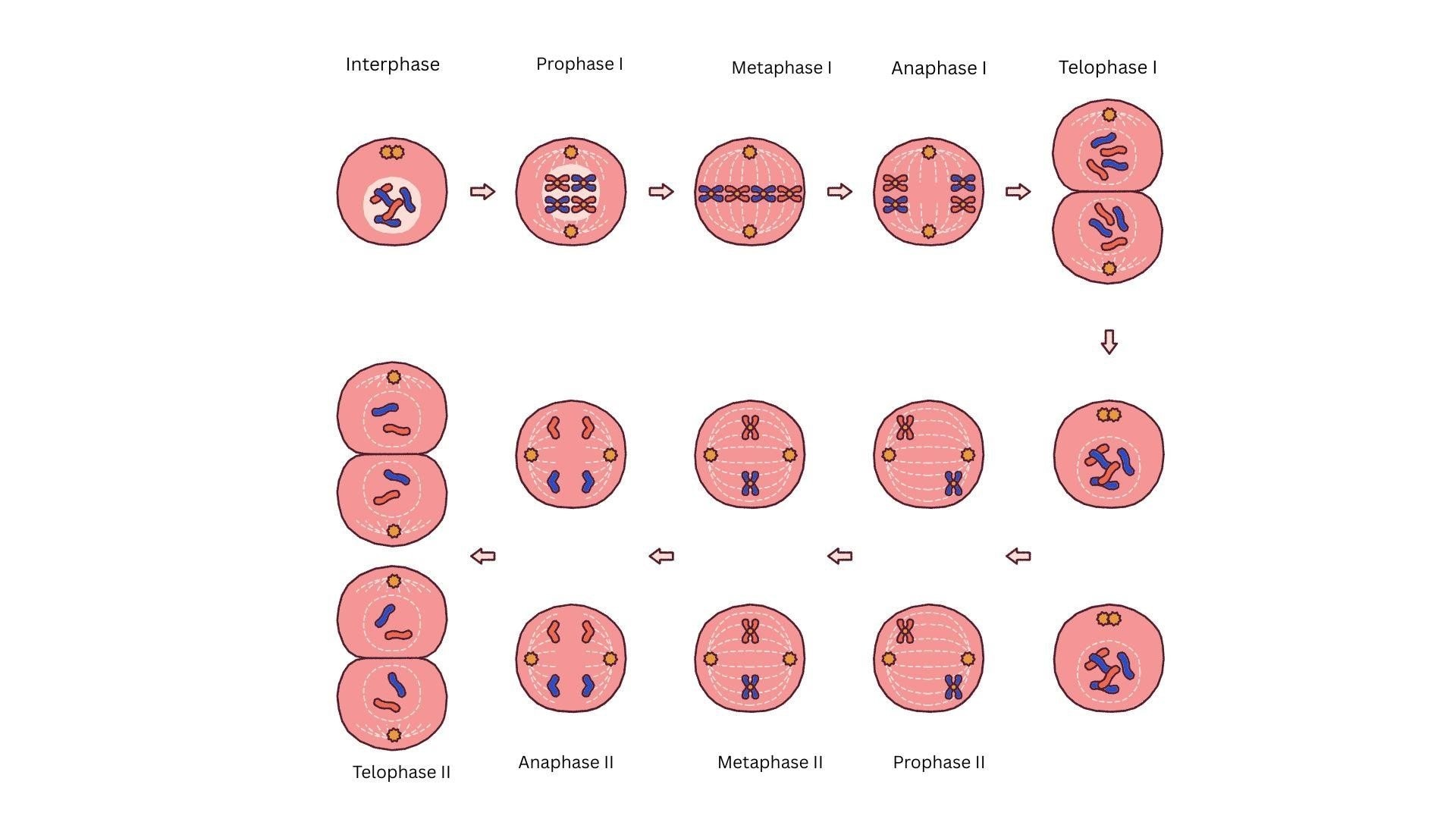
The 8 Stages of Meiosis
Both Meiosis I and Meiosis II have four stages: Prophase, Metaphase, Anaphase, and Telophase. Before the stages of Meiosis I, interphase prepares the cell for division by promoting growth, DNA replication, and the assembly of the cell machinery necessary for the two cell divisions. Here are the stages of Meiosis I:
Prophase I: Crossing Over and Genetic Recombination
The largest phase of meiosis I is prophase I. It is further divided into 5 small phases: leptotene, zygotene, pachytene, diplotene, and diakinesis.
Leptotene: In leptotene, the loose and uncondensed chromatin starts to condense. It forms compact structures called chromosomes.
Zygotene: The pairing of homologous chromosomes starts in zygotene. This process is known as synapsis. During synapsis, a complex protein structure is formed between two homologous chromosomes. This structure is called the synaptonemal complex. It acts as glue to maintain the pairing of chromosomes. Pairing is known as a bivalency because every complex contains two chromosomes. A bivalve contains four chromatids. Therefore, it is also called a tetrad.
Pachytene: Chromatids of these homologous chromosomes form recombination nodules. Genetic material exchange between them. This process is known as crossing over. Crossing over is an enzyme-mediated process. The enzyme that is involved in the process of crossing over is called recombinase.
Diplotene: The separation of homologous chromosomes begins in diplotene. The synaptonemal complex dissolves there. During the Diplotene stage, homologous chromosomes begin to separate as the synaptonemal complex dissolves. They don't separate and remain attached at the site of crossing over. These X-shaped structures are called Chiasmata.
Diakinesis: Terminalization of chiasmata occurs during this process. Chromosomes undergo further condensation during this stage. At the end of diakinesis, the nucleolus disappears, and the nuclear envelope breaks down. Chromosomes prepare to align, and the meiotic spindle starts to assemble.
Metaphase I: Independent assortment of Chromosomes
In metaphase I, spindle fibers are attached to kinetochores of homologous chromosomes. Chromosomes align at the center of the cell to form the metaphase plate. This random alignment is the basis for the independent assortment of chromosomes, ensuring that each gamete receives a unique genetic combination.
Anaphase I and Telophase I: First Division Complete
- In anaphase I, homologous chromosomes separate and move toward opposite poles.
- During telophase, the nuclear membrane and nucleolus reappear in the haploid set of chromosomes.
- After the formation of two haploid daughter cells, the division of cytoplasm occurs, which is known as cytokinesis.
- Each daughter cell contains one chromosome from each pair, and each chromosome contains two sister chromatids.
Meiosis II: Creating Four Haploid Gametes
Meiosis II starts with 2 haploid daughter cells formed at the end of Meiosis I. It produces 4 haploid gametes by chromatid separation, like mitosis
- Prophase II: In prophase II, nuclear membranes start to disappear, and chromosomes become compact.
- Metaphase II: Chromosomes align at the cell's equator. And spindle fibers attach to the kinetochores.
- Anaphase II: The centromere splits in anaphase II. Sister chromatids pull toward opposite sides of the cell.
- Telophase II, Cytokinesis and formation of 4 haploid gametes: chromosomes reach the poles and start to decondense, and the nuclear envelope reforms in telophase II. After that, cytokinesis occurs to produce 4 haploid gametes.
How Does Meiosis Differ from Mitosis?
Meiosis and Mitosis are two forms of cell division. They serve different purposes. Meiosis occurs in germ cells and produces haploid gametes that are necessary for sexual reproduction. Mitosis occurs in somatic cells of the body and is crucial for the healing, repairing, or growth of an organism.
Chromosome Number: Diploid vs Haploid Cells
In Mitosis, a diploid parent cell (2n) produces two identical daughter cells. Each of them has the same number of chromosomes. This form of cell division is necessary for the normal functions of the body, such as growth and repair of body parts.
In contrast, the number of chromosomes is reduced to half in Meiosis from diploid cells (2n) to haploid (n) gametes. They receive half the genetic material of the parent cell. It is necessary for reproduction and genetic diversity.
Cell Division Types: When Each Process Occurs
Both Mitosis and meiosis occur at different times in different cells of the body. They carry out their respective functions.
- Mitosis occurs in somatic cells or body cells and continues throughout the life span of an individual. It works by producing two identical diploid daughter cells. It is responsible for wound healing, the replacement of damaged cells, and body growth.
- Meiosis occurs only in specialized cells called germ cells, which are present in the gonads or reproductive organs of an individual. It produces 4 genetically different haploid cells or gametes in two stages. It contributes to genetic variation by producing genetically unique offspring from gametes.
How to Draw a Meiosis Diagram: Step-by-Step Tutorial
Drawing a Meiosis diagram can be interesting if the right tools are used. In this digital age, we can draw diagrams on digital tools like EdrawMax. Whether you are worried about your assignment or presentation, or want to teach meiosis to students, a Meiosis drawing will not be a problem. Here is a step-by-step guide to drawing a meiosis diagram:
Drawing Meiosis I with Template
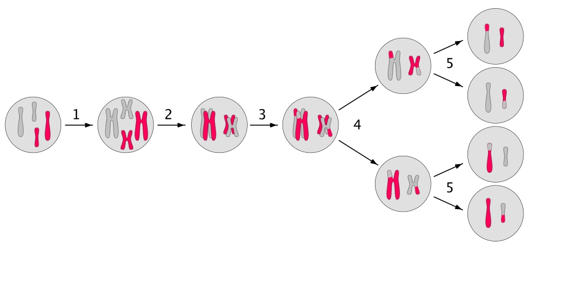
Draw two horizontal columns for Meiosis I and Meiosis II. To draw a Meiosis I diagram, make shapes for all the stages
- In the first oval shape, draw a diploid germ cell after DNA replication and label it. Now, draw paired X-shaped chromosomes to represent homologous chromosomes.
- For Prophase I, draw an oval shape and align these chromosomes as tetrads. Show and label chromosome crossing.
- Draw a shape with homologous chromosomes aligning with the metaphase plate in Metaphase I. Label the independent assortment of chromosomes.
- Now, show the chromosomes pulling to the opposite poles in Anaphase I (while keeping the X shape intact). Label reductional division
- Now draw two circles for daughter cells with partially divided cytoplasm for Telophase I. Then, separately draw two circles for the cytokinesis stage. Label these two daughter cells as haploid cells.
Make a Biology Diagram Now for Free with EdrawMax
Although creating a clear and well-labeled meiosis diagram can be difficult, an easy-to-use diagramming tool could lead to a flat learning curve. Now, if you are looking for a tool that a beginner would find simple to start with, EdrawMax is what you need. Follow these simple steps below to make a biology diagram on EdrawMax for free:
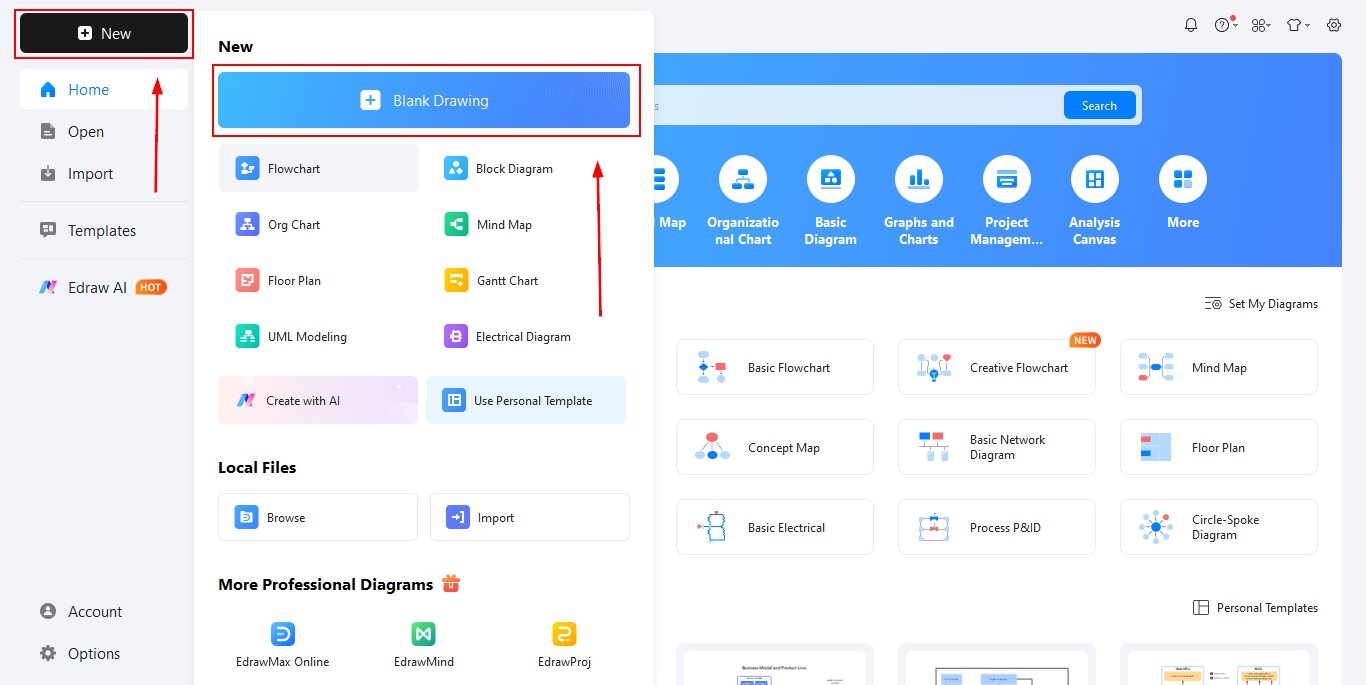
Step1 Open the tool and get started
Launch EdrawMax on your computer and log in or continue as a guest. Create a new project by selecting a blank canvas or choosing a biology-related template to begin your meiosis diagram.
Set up the workspace by adjusting the page size, orientation, and background to ensure enough space for all meiosis stages to be displayed clearly and neatly.
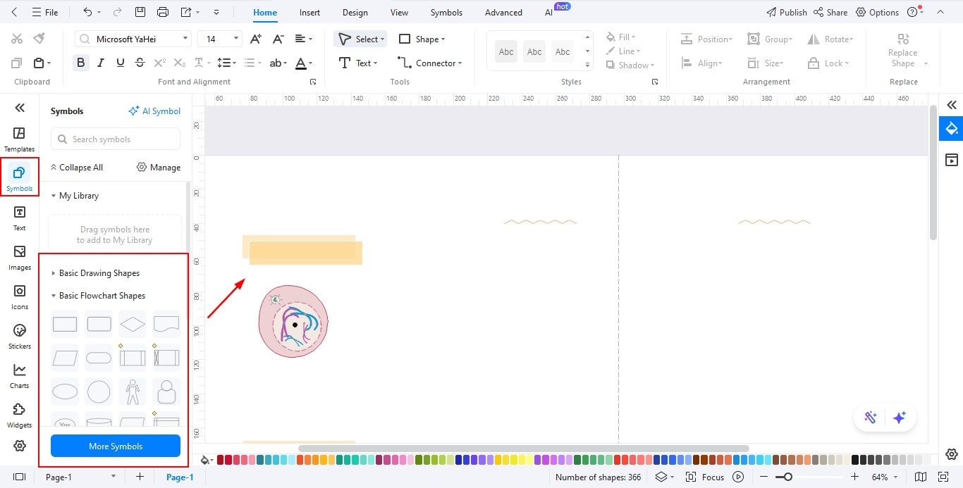
Step2 Drag all elements needed
Open the symbol library and search for shapes representing cells, nuclei, chromosomes, and other biological components required for meiosis.
Drag and drop these elements onto the canvas, duplicating them as needed to represent different phases such as Prophase, Metaphase, Anaphase, and Telophase.
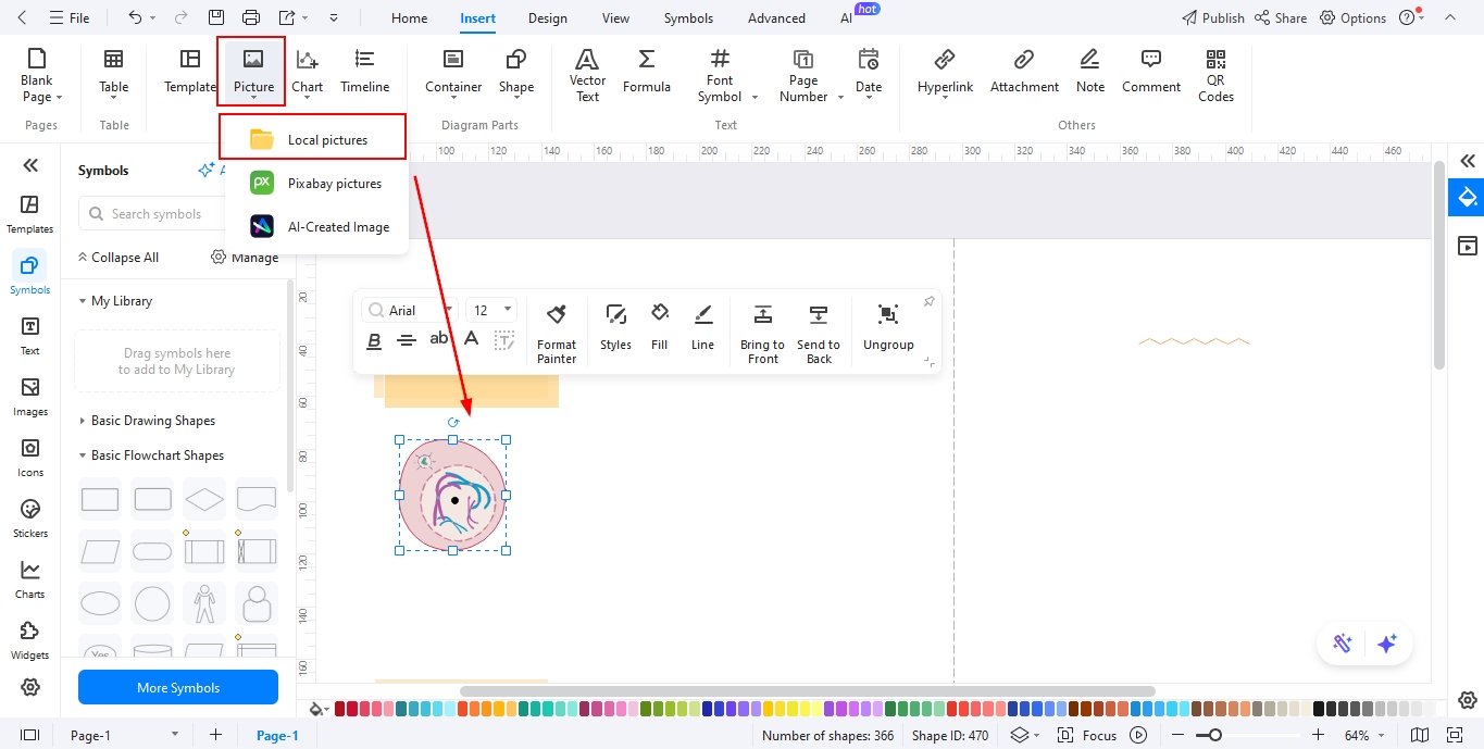
Step3 Add images to the diagram
Insert high-quality images or illustrations for Meiosis I and Meiosis II using the Insert Image option to enhance visual understanding.
Resize, rotate, and position each image properly so that the stages appear organized and visually balanced across the diagram.
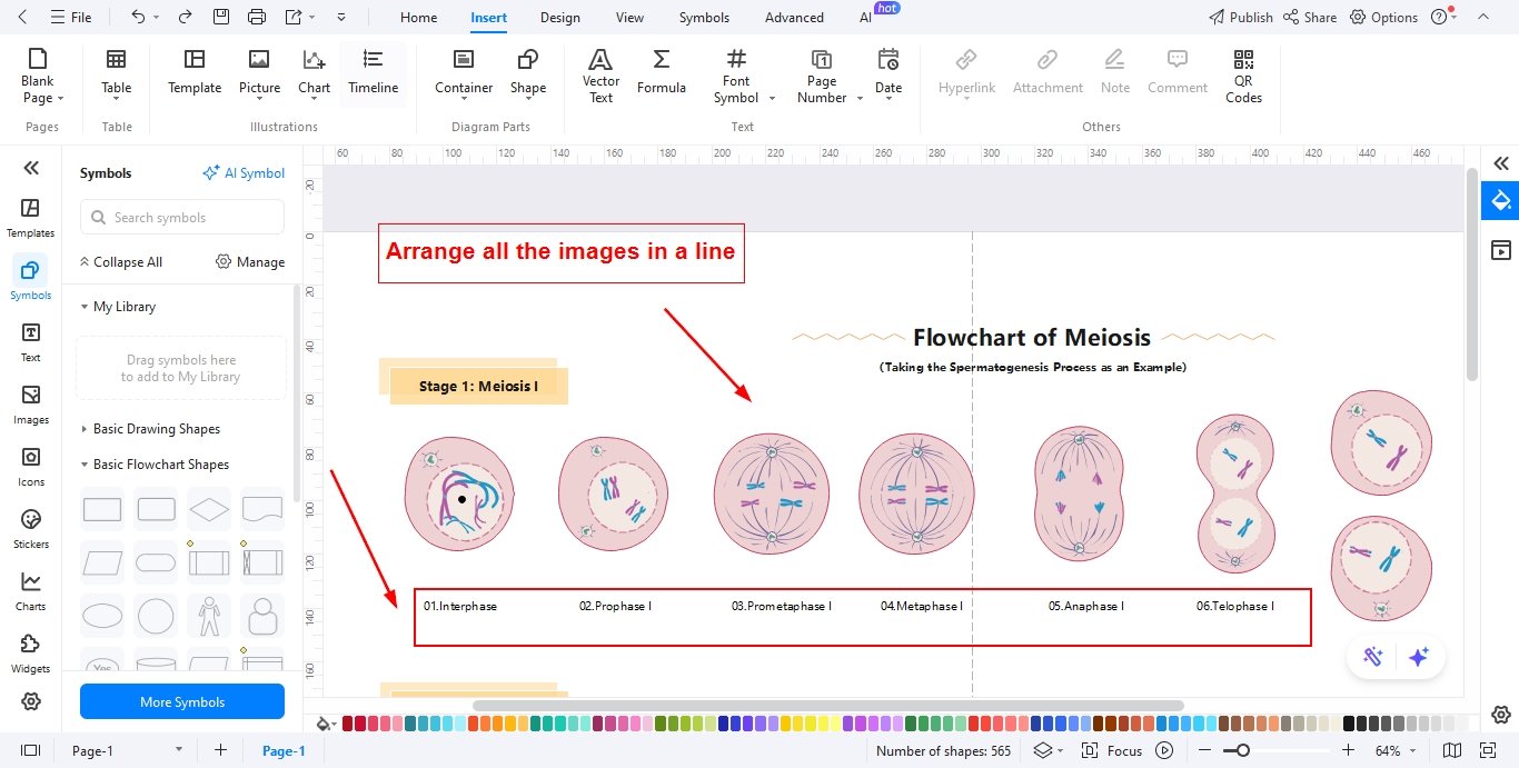
Step4 Add text for each image and arrange them in line
Use the text tool to label every phase clearly, including stage names and short descriptions explaining key events.
Align all images and text boxes in a straight line or structured flow to show the correct sequence of meiosis progression.
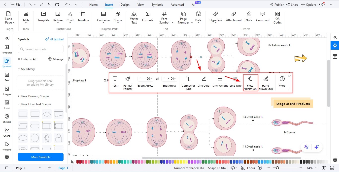
Step5 Add relationship among the phases using the connector tool
Use arrows or connectors to link one phase to the next, visually representing the continuous process of cell division.
Customize connector styles, arrowheads, or colors to make relationships between stages clear and easy to follow.
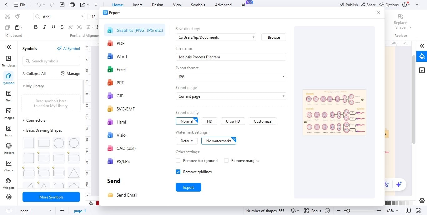
Step6 Export the diagram
Review the entire diagram for accuracy, clarity, and alignment before exporting. Make sure all labels and connections are readable.
Export the final diagram in your preferred format, such as PDF, PNG, or JPG, for assignments, presentations, or printing.
Bonus: Tips for Visualizing Crossing Over and Genetic Recombination
Crossing over and genetic recombination are important features of Meiosis I. Most students find it difficult to visualize them both on digital tools and with their hands.
Crossing over visualization techniques for drawing Meiosis on digital tools:
- Use frame-by-frame animations, e.g., on PowerPoint, Canva, or EdrawMax, etc., for tetrad formation and crossing over.
- Use different colors for chromosomes so that genetic recombination can be shown properly
- Use clickable, separate layers:
- For a base consisting of homologous chromosomes
- Crossing over of genetic material
- Third for labeling
Crossing over visualization techniques for drawing Meiosis on the classroom board:
- Use small pieces of colored pipe cleaners or yarns to show chromatids and beads to show the gene locus.
- You can use paper strips of different colors to visualize recombinant genetic material.
- Similarly, colored chalks or board markers can be used to draw chromatids. Exchanged portions can be erased or redrawn.
FAQs About Meiosis
-
Why Do We Need Two Divisions in Meiosis?
Meiosis completes in two successive divisions to produce 4 haploid gametes because it has two goals:
- Reduction of chromosome number and exchange of genetic material (Meiosis I):
In this division, the chromosome number is reduced to half from diploid (2n) to haploid (n). Genetic material is exchanged between homologous chromosomes through crossing over. It is crucial for genetic variations in the next generation. Homologous chromosomes are separate, and 2 haploid daughter cells are formed at the end of this division.
- Separation of sister chromatids (Meiosis II):
During this division, sister chromatids separate, and 4 haploid cells are formed. These haploid cells make a diploid zygote upon fertilization. Without Meiosis II, each gamete would have a duplicated chromosome. It could result in genetic disorders in offspring.
So, two divisions are necessary for the exchange of genetic material and the formation of 4 haploid gametes. They ensure genetic diversity and the correct number of chromosomes in each generation.
- Reduction of chromosome number and exchange of genetic material (Meiosis I):
-
How Many Chromosomes Are There in Each Gamete?
In all species, the gametes carry half the number of chromosomes as compared to somatic cells. For example, in humans, Somatic cells or diploid cells have 46 chromosomes (23 pairs) while Gametes are haploid and carry 23 chromosomes, which is half the number of the parent cell.
Similarly, cats have 38 chromosomes, and their gametes have 19 chromosomes.
The reduction in the number of chromosomes ensures the diploid number of chromosomes in an offspring by the fusion of two haploid gametes.
AI Diagram Generator
Enter your prompt. Upload files if needed. Generate diagrams, charts, or slides instantly.