About this eye diagram template
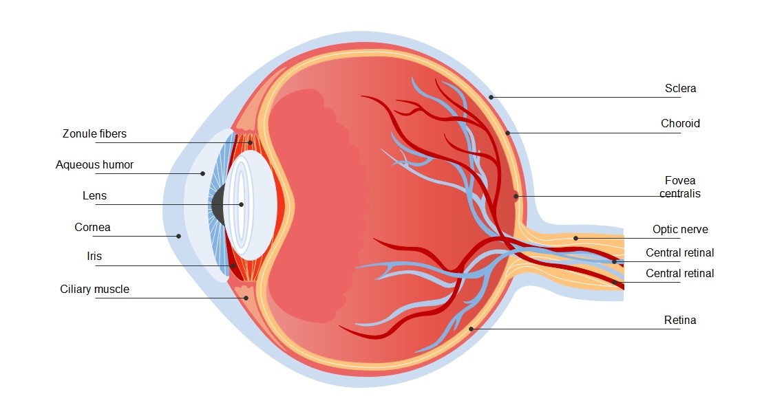
This eye diagram template provides a high-quality cross-section of the human eyeball. It is designed to help students and educators visualize internal structures easily. Use this resource to study how light moves from the cornea to the retina to create the sense of sight.
Frontal Structures
The front section of the eye is responsible for focusing and protecting the internal environment. These parts capture incoming light and adjust it so that a clear image can form on the light-sensitive back layer.
- Cornea: The clear outer window that focuses light.
- Iris: The colored muscle that controls the pupil size.
- Lens: A flexible structure that fine-tunes image focus.
- Aqueous humor: Watery fluid that provides nutrients and pressure.
- Ciliary muscle: The muscle that changes the lens shape.
- Zonule fibers: Fibers that hold the lens in place.
Inner and Rear Anatomy
The back of the eye processes light into electrical signals sent to the brain. These tissues and nerves are critical for detail, color, and depth perception. They ensure that we can see clearly in various lighting conditions.
- Retina: The nerve layer that senses light and images.
- Optic nerve: The cable that sends signals to the brain.
- Sclera: The white protective outer coat of the eye.
- Choroid: The vascular layer providing blood to the retina.
- Fovea centralis: The spot responsible for sharp, central vision.
- Central retinal vessels: Vessels supplying blood to the inner eye.
FAQs about this Template
-
What is the primary role of the cornea in vision?
The cornea acts as the eye's outermost lens. It functions like a window that controls and focuses the entry of light into the eye. Because it is curved, it helps bend light so it can land perfectly on the lens. It also protects the inner parts of the eye from dust, germs, and other potentially harmful external particles.
-
How does the iris control the amount of light entering the eye?
The iris is the colored part of your eye containing muscles that expand or contract. It regulates the size of the pupil, which is the opening in the center. In bright light, the iris closes the pupil to protect the retina. In low light, it opens the pupil wider to allow more light in, helping you see better in the dark.
-
Why is the optic nerve considered a vital part of the eye?
The optic nerve is a vital communication cable between the eye and the brain. It is made up of millions of nerve fibers that carry electrical impulses from the retina. Without this nerve, the brain would never receive the visual information captured by the eye. It is the final step in the vision process, allowing us to perceive our surroundings.