About this heart valve diagram template
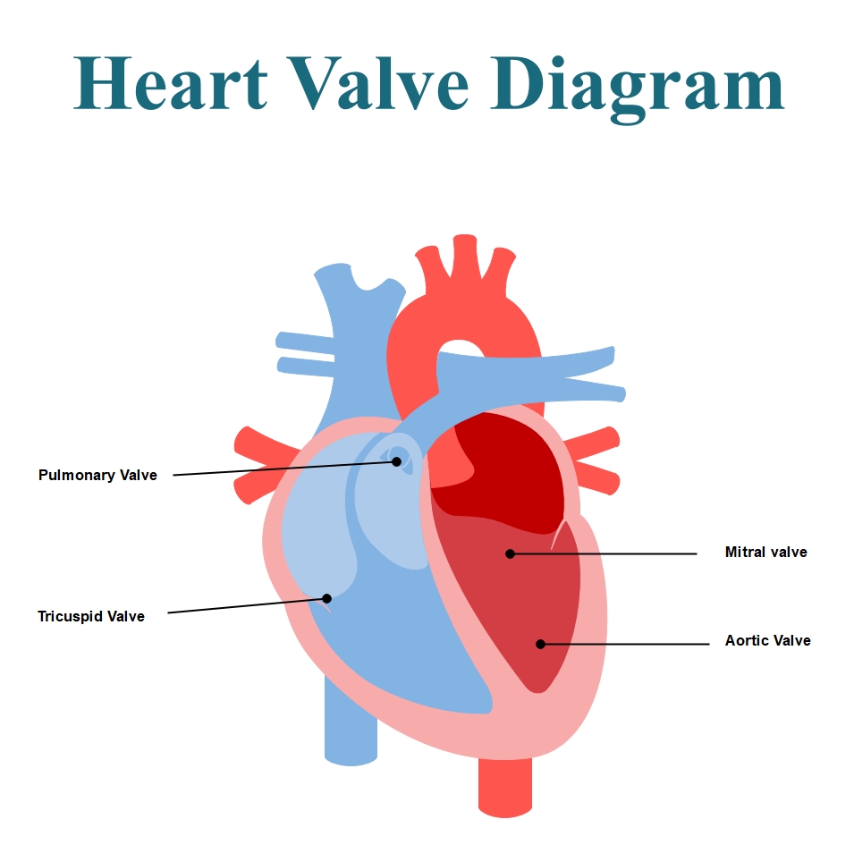
This diagram provides a clear visual representation of the internal structures of the human heart. It highlights the four essential valves that regulate blood flow. Use this template for medical education, school projects, or healthcare presentations to simplify complex anatomy.
Atrioventricular Valves
The atrioventricular valves separate the heart's upper atria from the lower ventricles. They prevent blood from flowing backward when the heart chambers contract. These valves are critical for ensuring efficient movement of blood through the heart.
- Tricuspid Valve: This valve is located between the right atrium and the right ventricle.
- Mitral Valve: This valve is positioned between the left atrium and the left ventricle.
Semilunar Valves
Semilunar valves are found at the bases of the large arteries leaving the heart. They control the exit of blood from the ventricles into the circulatory system. Their unique shape allows them to close tightly after blood passes.
- Pulmonary Valve: This valve regulates blood flow from the right ventricle into the lungs.
- Aortic Valve: This valve controls the flow of oxygen-rich blood from the left ventricle.
FAQs about this Template
-
What is the primary function of heart valves?
Heart valves act like one-way doors to ensure blood flows in the right direction. They open to let blood pass and close tightly to prevent any backflow. This mechanism is crucial for maintaining proper blood pressure and oxygen delivery throughout the body. If a valve fails to close properly, it can lead to serious health complications or heart failure.
-
Which heart valve is most commonly affected by disease?
The mitral valve is frequently associated with medical conditions such as mitral valve prolapse or regurgitation. Because the left side of the heart works harder to pump blood to the rest of the body, these valves endure higher pressure. Regular check-ups are important because early detection of valve issues can prevent long-term damage to the heart muscle.
-
How does a heart valve diagram help in medical education?
A visual diagram simplifies the complex anatomy of the cardiovascular system for students and patients. By labeling each valve, people can better understand the pathway of blood from the lungs to the body. Diagrams serve as an effective communication tool in clinical settings. They help doctors explain specific conditions or surgical procedures to their patients clearly.