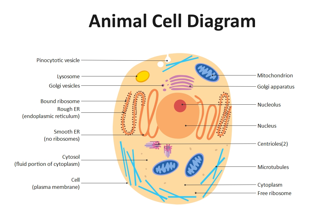
About this Animal Cell Diagram Animated template
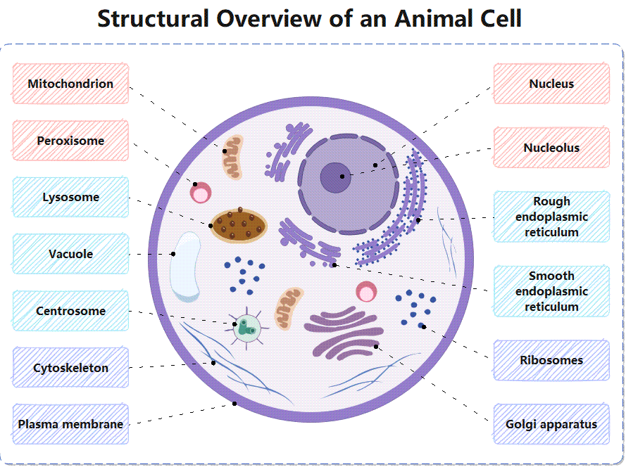
This template provides a clear visual breakdown of an animal cell. It labels every major organelle, from the nucleus to the plasma membrane. It is perfect for biology presentations, classroom posters, or digital learning materials to help students grasp cellular structures quickly.
Genetic Control Center Organelles
This section focuses on the cell's command center. The nucleus acts as the primary brain, while the nucleolus produces ribosomes. Together, they manage growth and reproduction by housing DNA and providing essential instructions for life.
- Nucleus
- Nucleolus
Energy and Protein Manufacturing
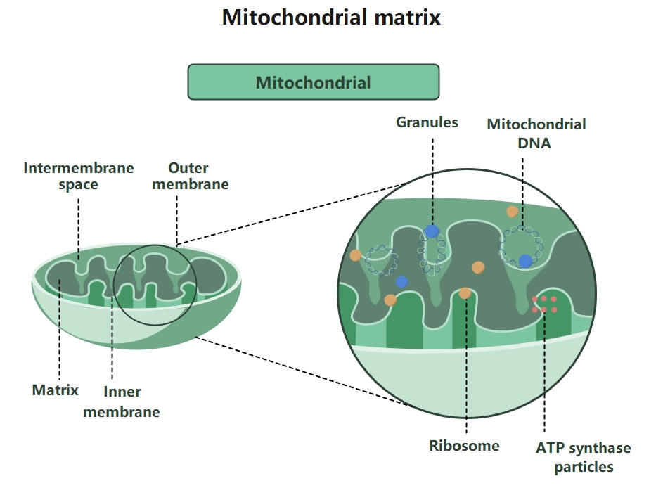
These parts handle energy production and molecule building. Mitochondria generate power through respiration. Ribosomes and endoplasmic reticulum work together to build proteins. These components are vital for maintaining the cell's daily activity and physical health.
- Mitochondrion
- Ribosomes
- Rough endoplasmic reticulum
- Smooth endoplasmic reticulum
Cellular Storage and Waste
Cells need efficient ways to store nutrients and remove harmful waste. Lysosomes digest materials, while peroxisomes break down toxins. Vacuoles store water and food, ensuring the cell remains clean and balanced throughout its entire life cycle.
- Lysosome
- Peroxisome
- Vacuole
Structural Support and Transport
These components provide the cell's shape and move materials internally. The Golgi apparatus packages proteins for transport. The cytoskeleton and plasma membrane provide physical support, while centrosomes assist in the critical process of cellular division.
- Golgi apparatus
- Cytoskeleton
- Plasma membrane
- Centrosome
FAQs about this Template
-
What are the most important organelles in an animal cell?
The most important organelles include the nucleus, which acts as the control center, and the mitochondria, which provide energy. Additionally, the ribosomes and endoplasmic reticulum are crucial for protein synthesis. While every part plays a role, these core components manage the genetic information and energy flow necessary for the cell to stay alive and function properly.
-
How does an animated diagram improve biology learning?
Animated diagrams are effective because they make abstract biological concepts much more concrete. By showing the spatial relationships between organelles like the Golgi apparatus and the nucleus, students can better understand how cells work. Visual aids help with memory retention and keep learners engaged, making complex scientific topics like cellular biology feel much more accessible and interesting.
-
What is the difference between smooth and rough endoplasmic reticulum?
The primary difference between smooth and rough endoplasmic reticulum is the presence of ribosomes. Rough ER is covered in ribosomes, giving it a bumpy look and focusing on protein production. In contrast, smooth ER lacks these ribosomes and specializes in creating lipids and detoxifying chemicals. Both are essential for the cell's ability to manufacture and process different types of biological molecules.